NikonUSA NikonNet MicroscopyU NikonMall NikonSchool
Search
Go

Fluorescence Microscopy Digital Image Gallery

The widefield reflected light fluorescence microscope has been a fundamental tool for the examination of fluorescently labeled cells and tissues since the introduction of the dichromatic mirror in the late 1940s. Furthermore, advances in synthetic fluorophore design coupled to the vast array of commercially available primary and secondary antibodies have provided the biologist with a powerful arsenal in which to probe the minute structural details of living organisms with this technique. In the late twentieth century, the discovery and directed mutagenesis of fluorescent proteins added to the cadre of tools and created an avenue for scientists to probe the dynamics of living cells in culture. This gallery examines the fluorescence microscopy of both cells and tissues with a wide spectrum of fluorescent probes.

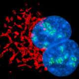
Animal and Human Cells in Culture - The fluorescence microscope provides an interesting window into the world of the cell and is one of the biologist's favorite tools for the examination of both living and fixed cells in culture. The ability to specifically target organelles and macromolecules with synthetic fluorophores and immunofluorescence has produced a virtual revolution in the dynamic field of fluorescence microscopy for the examination of cells in culture. This portion of the fluorescence digital image gallery features widefield fluorescence images captured from over 30 cell lines stained with a mixture of synthetic probes, antibodies, and fluorescent proteins.

Rat Tissue Sections - The humble rat has had an outsized impact on human history. In the Middle Ages, the black rat (Rattus rattus) was blamed for spreading the Black Plague through its fleas, a pandemic that killed a third of Europe's population, an estimated 34 million people. In modern times, however, a larger cousin, the Brown rat (Rattus norvegicus) has become an important model organism in biological research. Selective breeding of the Brown Rat has produced the albino laboratory rat. Rats grow quickly to sexual maturity and are easy to keep and breed in captivity. Scientists have bred many strains or "lines" of rats specifically for experimentation. Generally, these lines are not transgenic because the easy techniques of genetic transformation that work in mice do not work as well for rats. This has been a problem for investigators who view rat behavior and physiology as more relevant to humans and easier to observe than in mice. In October 2003, researchers succeeded in cloning two laboratory rats by the problematic technique of nuclear transfer. As cloning techniques are perfected, rats likely will become an important subject of genetic research.

Human Lung Tissue Sections - The lungs are the elastic respiratory organs of vertebrates that breathe air. In humans, the lungs are paired in the chest cavity on opposite sides of the heart. The right lung is divided into three lobes and the left is composed of two lobes. A membranous lining called the pleura covers the human lungs, as well as the upper body cavity. A small amount of watery fluid is generally present in the pleural cavity that keeps the pleura lubricated and allows the lungs to move smoothly in the chest cavity during the breathing process.

Human Prostate Gland Tissue Sections - The prostate is a key component of the male reproductive system located directly below the bladder where it encircles the upper portion of the urethra. The gland is extremely small at birth, but enlarges to about the size of a walnut during adolescence. The primary function of the prostate gland is to secrete prostatic fluid, which contains calcium, zinc, phosphate, citrate, and various enzymes. Prostatic fluid is contributed to the seminal fluids that carry sperm and is thought to be important for the successful fertilization of an egg.

Human Small Intestine Tissue Sections - In humans, the small intestine is generally more than 20 feet long, making it a much more prominent digestive structure than the small intestines of most nonhuman primates. This notable difference reflects the significant divergence in diet of omnivorous humans from the great apes, which primarily are vegetarians that consume large amounts of low-quality plants with a high proportion of roughage. The surface area of the lining of the human small intestine is approximately 300 square meters, an area roughly equivalent to that of a standard tennis court.

Human Thyroid Tissue Sections - The thyroid gland is a component of the endocrine system comprised of two oblong lobes linked by a narrow piece of tissue called the isthmus. The thyroid is located in the throat and contains millions of follicles that act as storage sites for the various hormones produced by the gland. Thyroid secretions function in a variety of ways in the human body but are particularly important in adults as regulators of metabolism. The hormones are also central to normal growth and development in children. A childhood deficiency in thyroid hormones has been linked to dwarfism and mental retardation.

Mouse Brain Tissue Sections - The mouse is widely utilized as an animal model of a variety of brain diseases, such as Alzheimer's disease, encephalitis, dementia, and Creutzfeldt-Jakob disease. This practice can lead to important developments in the understanding of brain diseases and in the advancement of potential treatments. In one recent study, for instance, scientists employing a mouse model of spinocerebellar ataxia 1 (SCA1) discovered an effective way to "silence" the mutant gene allele or variant that causes the disorder without affecting the normal gene allele. SCA1 is a hereditary progressive neurodegenerative disorder that induces ataxia and other movement problems as an increasing number of brain cells are affected. Mice in the investigation that received the experimental gene therapy, however, were successfully protected from the disease and did not suffer from any abnormality of movement. The study suggests that similar methods may someday prove useful in the treatment of human neurodegenerative disorders.

Mouse Kidney Tissue Sections - Kidneys are paired organs that function chiefly in the filtration of the blood supply, maintenance of water balance, and expulsion of waste products. Mammalian kidneys are composed of numerous small lobules organized into an outer cortex and an inner medulla. Nephrons are the basic functional units of the kidneys and are responsible for the production of urine as they remove unneeded materials from the blood. In mammals, nephrons are long narrow tubes that are closed and folded at one end into a structure known as Bowman’s capsule that encases a group of capillaries collectively termed the glomerulus.

Mouse Intestine Tissue Sections - The intestines are considered to be divided into several different regions that extend from the stomach to the anus. The most basic division of the intestines is into the small intestine and the large intestine. The small intestine is the section closest to the stomach and is generally much longer and narrower than the large intestine. A variety of afflictions can affect the small and large intestines, such as enteritis, colitis, diverticulosis, and cancer. Various strains of laboratory mice are often utilized as models for such conditions.

Sheep Bladder Tissue Sections - The bladder is a collapsible, thick-walled muscular sac surrounded by a layer of fat that serves as a storage site for the urine produced by the kidneys. Urine is transported to the bladder via the ureters, each of which is linked to a kidney, and exits through the urethra. Depending on how much fluid it contains, the bladder may be expanded or contracted. Receptors located inside of the bladder indicate when the organ is sufficiently distended and activate a reflex pattern that causes the contraction of the organ's muscular wall and the relaxation of the internal urinary sphincter, allowing urine to enter the upper portion of the urethra.

Sheep Lung Tissue Sections - Sheep and other animal models are commonly employed to characterize the progression of human diseases and to test potential treatments. One notable recent study utilizing sheep focused upon using an artificial implantable lung as a temporary solution for lung failure. The pre-clinical testing of the device developed by researchers at the University of Michigan, which utilizes minute hollow fibers containing numerous micropores and the pumping force of the heart to oxygenate blood, was generally considered very promising. If further testing of the new artificial lung with sheep, and eventually human subjects, is similarly successful, the device may someday find use among patients on lung transplant waiting lists.

Sheep Tongue Tissue Sections - The tongue is a mass of striated muscle tissue intermingled with glands and fat that is encased in a mucus membrane and located along the floor of the mouth. The upper surface of the tongue has a rough appearance due to the presence of many minute projections termed papillae that contain the taste buds. Most vertebrates possess tongues, but many of the characteristics and functions of the organ vary. The long, flexible tongues belonging to anteaters and lizards, for instance, are important for capturing prey, whereas the tongue of a dog is critical in heat regulation and cats commonly use their tongues for grooming.

BACK TO FLUORESCENCE MICROSCOPY


Contributing Authors

Shannon H. Neaves, Douglas W. Martin, John D. Griffin, Nathan S. Claxton, Aferdita Ishmaku, J. David Homan, Richard L. Ludlow, and Michael W. Davidson - National High Magnetic Field Laboratory, 1800 East Paul Dirac Dr., The Florida State University, Tallahassee, Florida, 32310.