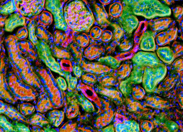
Mouse Kidney Tissue
The mouse kidney tissue sample presented in the digital image above was triple labeled before imaging. Alexa Fluor 568 (red fluorescence emission) conjugated to phalloidin was utilized to target polymerized actin (F-actin) and Alexa Fluor 488 (green emission) conjugated to wheat germ agglutinin was used to target sialic acid and N-acetylglucosaminyl residues. The popular nuclear and chromosome counterstain DAPI, which emits blue fluorescence upon binding to AT regions of DNA, was also employed. Images were recorded in grayscale with a 12-bit digital camera coupled to either a Nikon E-600 or Eclipse 80i microscope equipped with bandpass emission fluorescence filter optical blocks. During the processing stage, individual image channels were pseudocolored with RGB values corresponding to each of the fluorophore emission spectral profiles.
Featured in:
Share this page:


