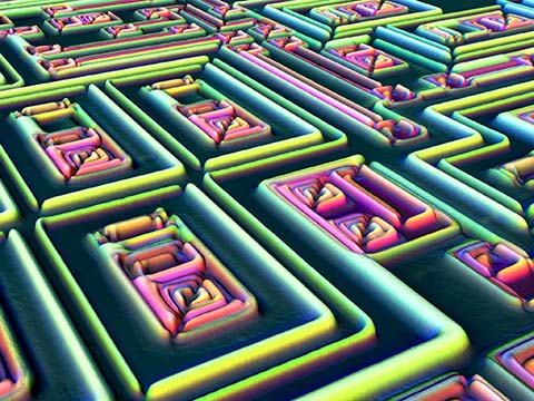
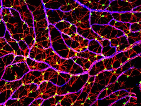
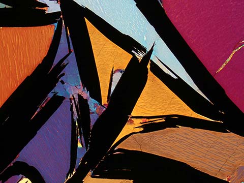
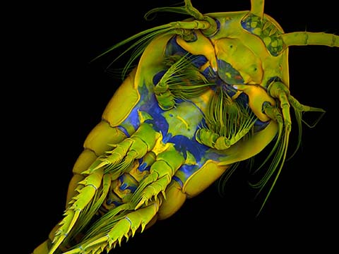
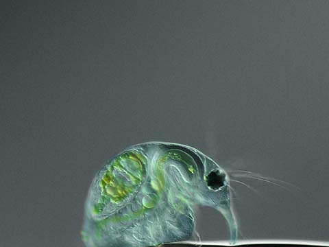
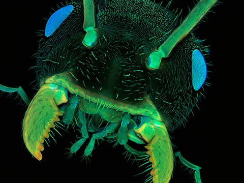
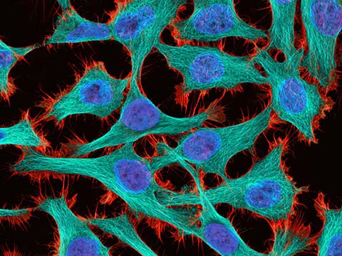
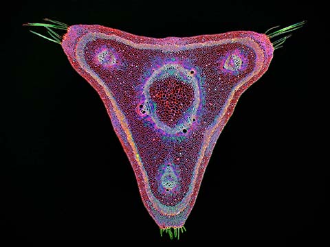
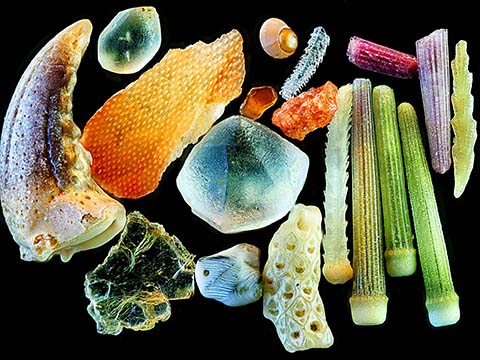
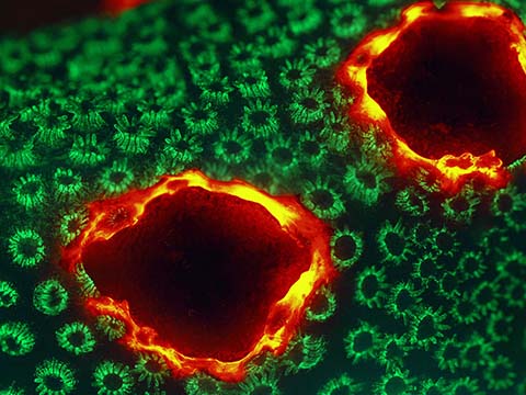
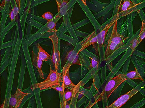
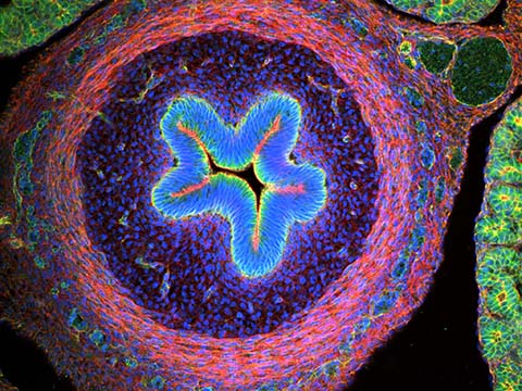
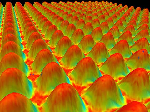
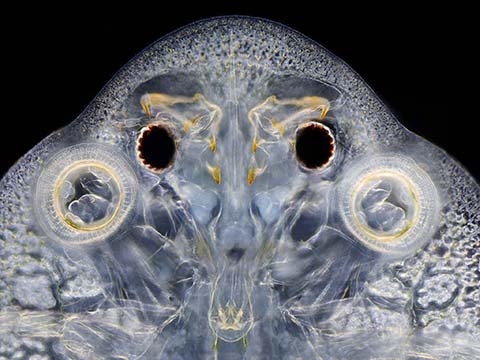
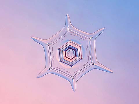
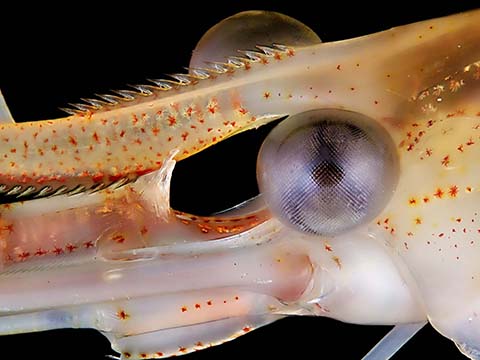
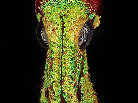
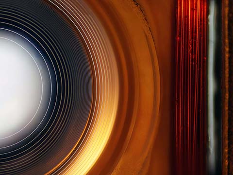
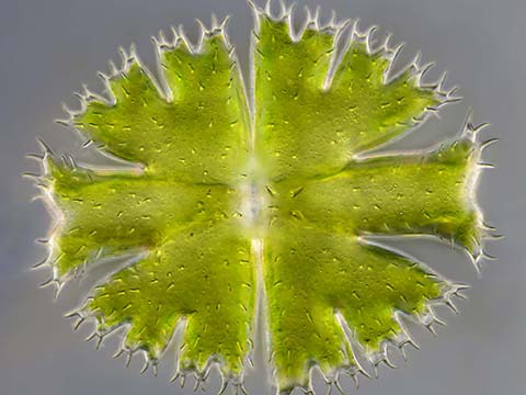
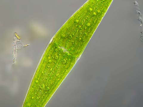
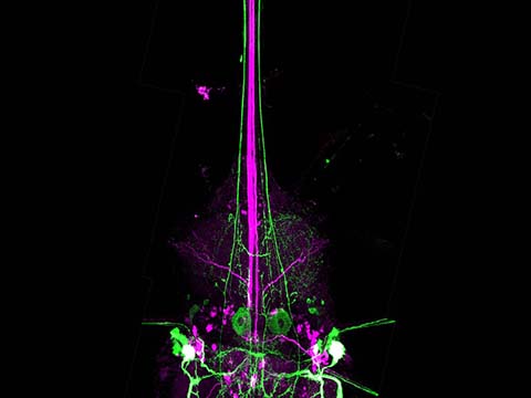
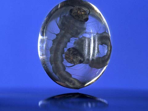
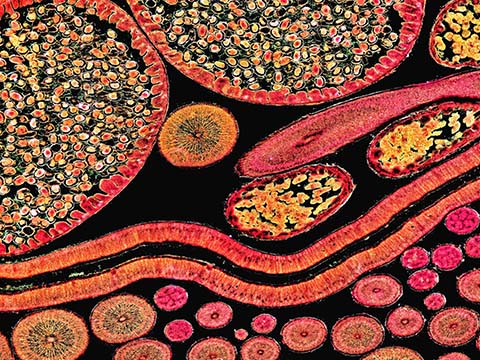
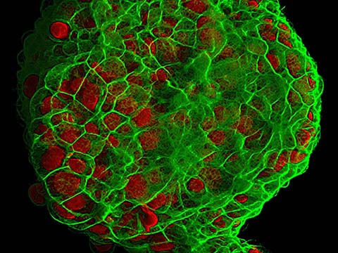
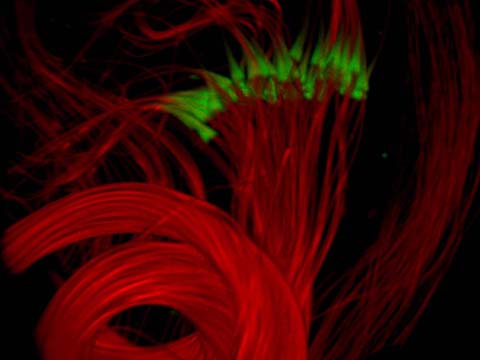
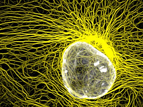
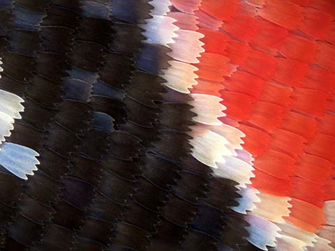
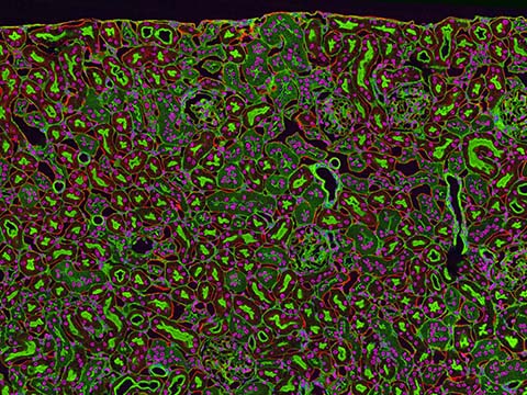
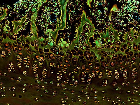
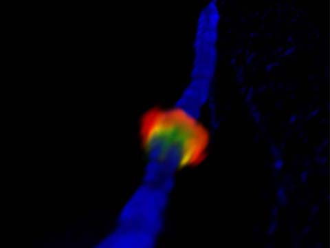
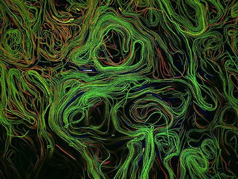
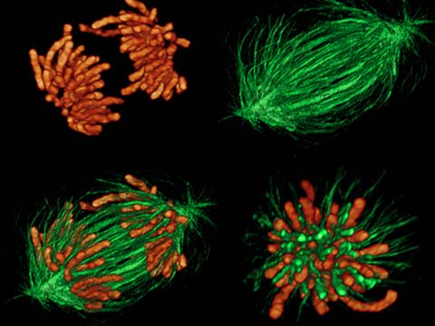
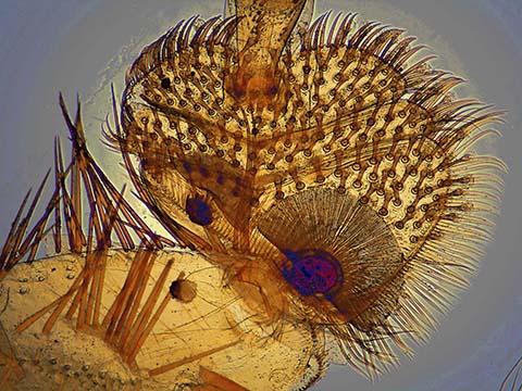
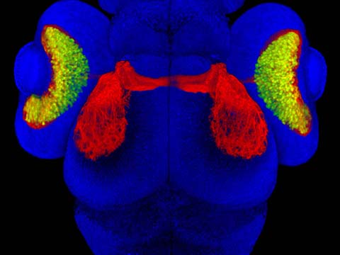
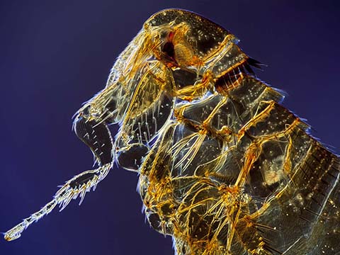
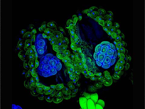
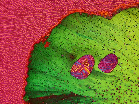
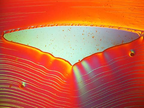
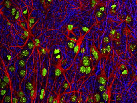
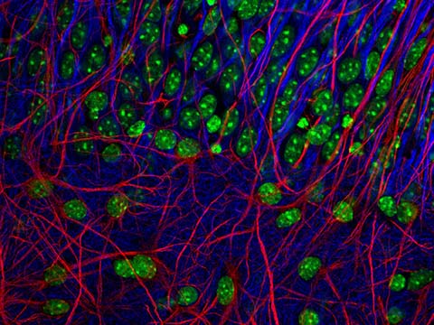
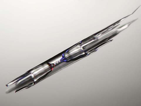
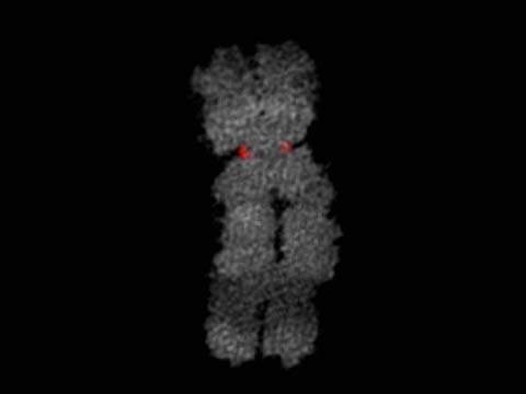
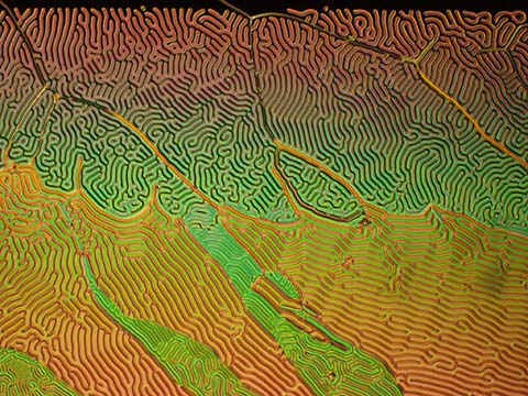
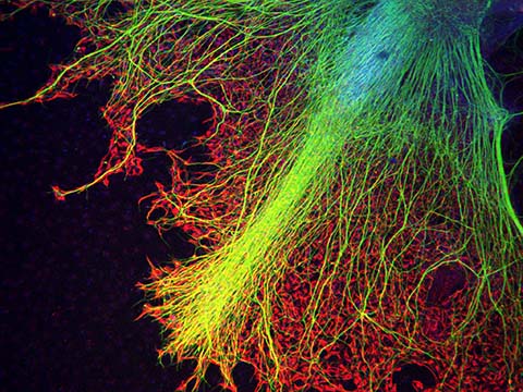
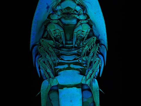
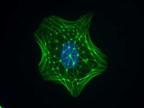
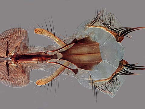
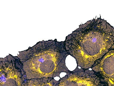
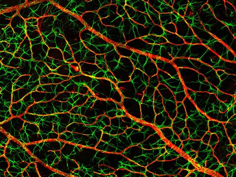
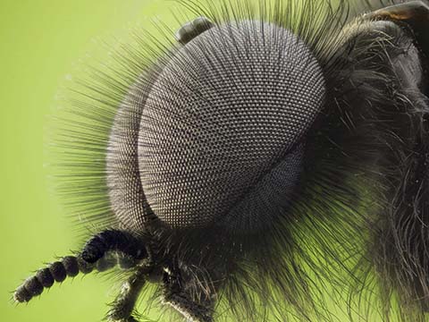
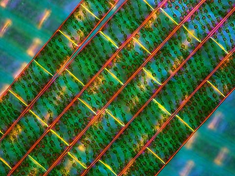
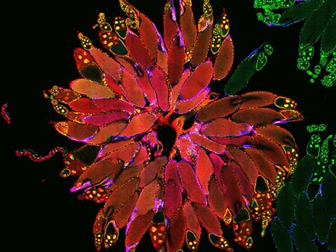
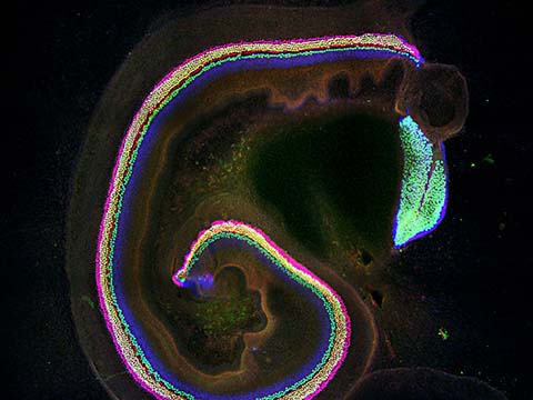
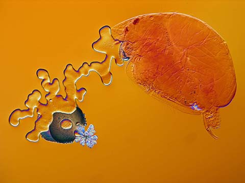
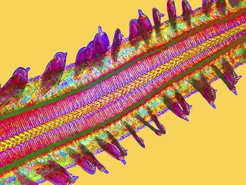
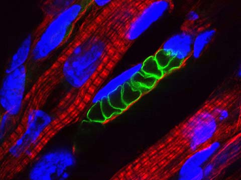
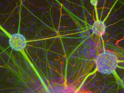
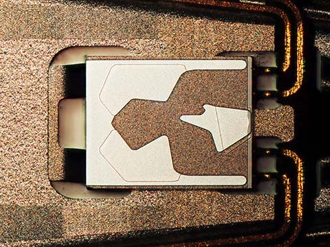
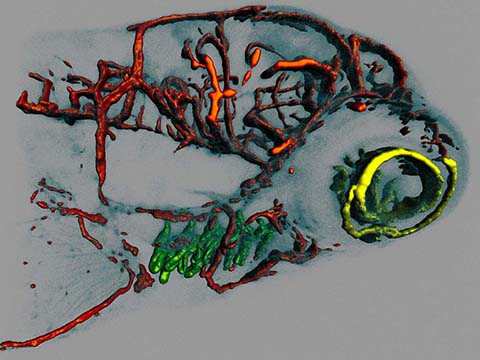
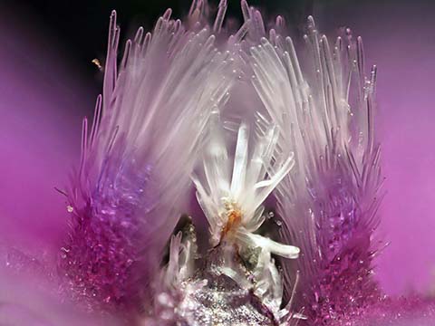
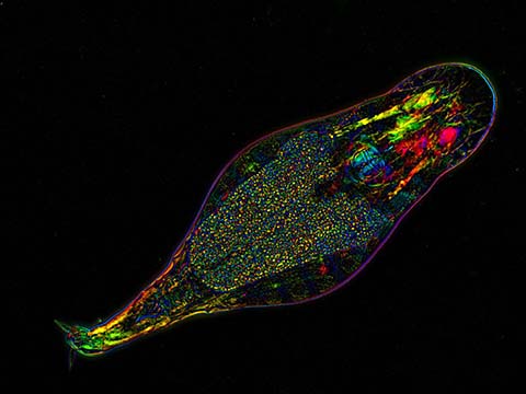
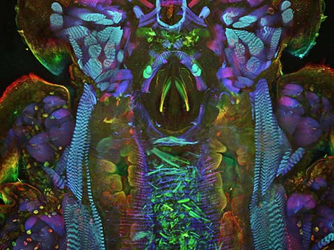
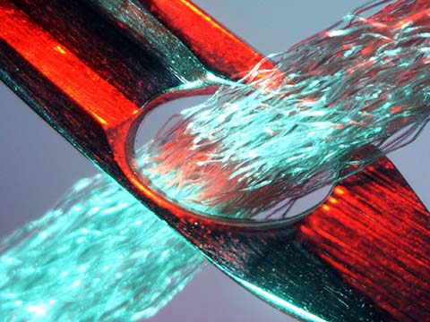
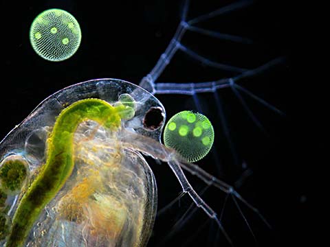
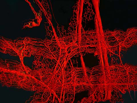
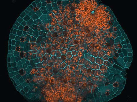
2011 Photomicrography Competition
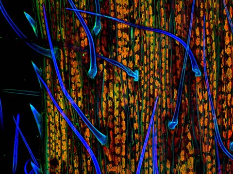
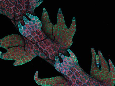
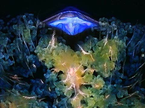
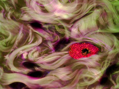
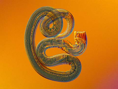
Top 20
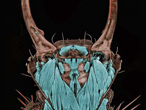
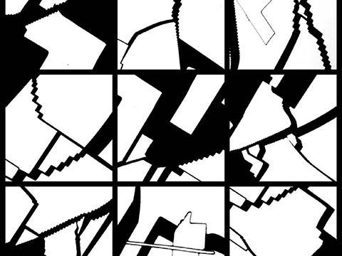

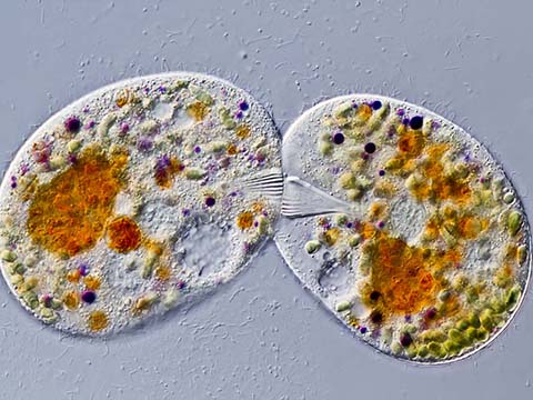
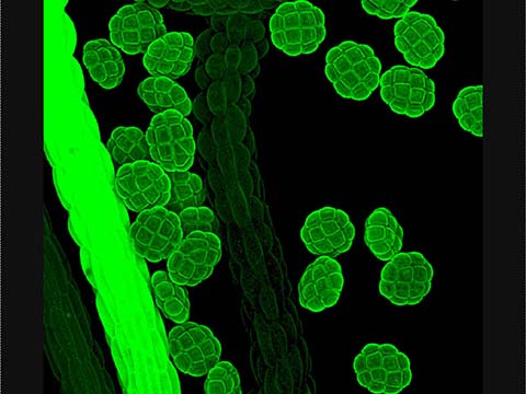
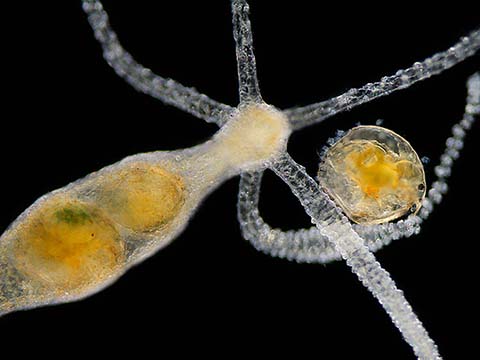
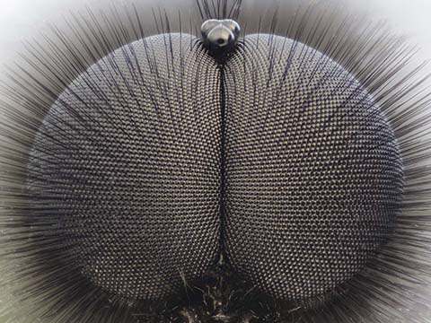
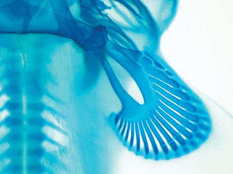
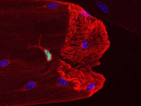
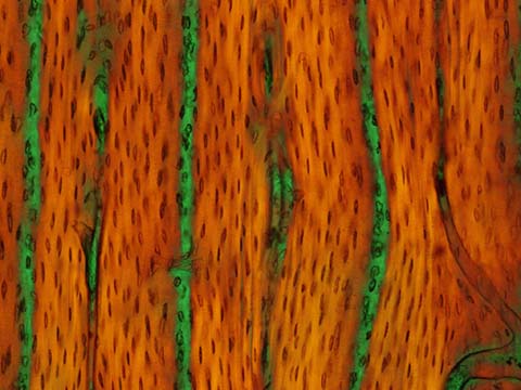
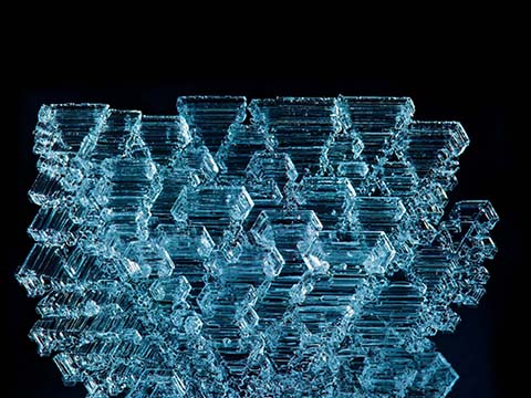
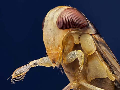
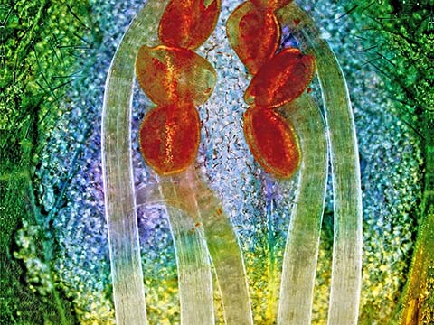
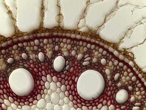
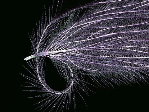
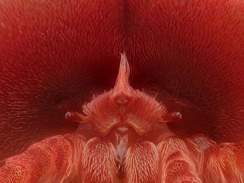
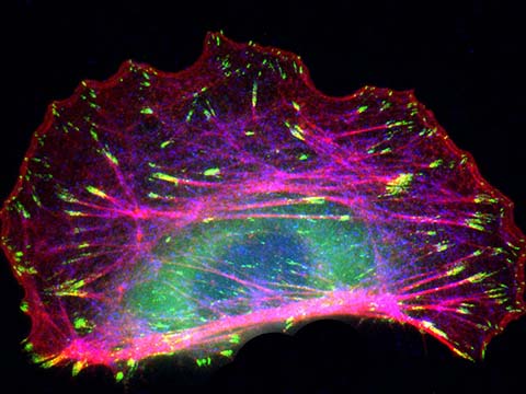
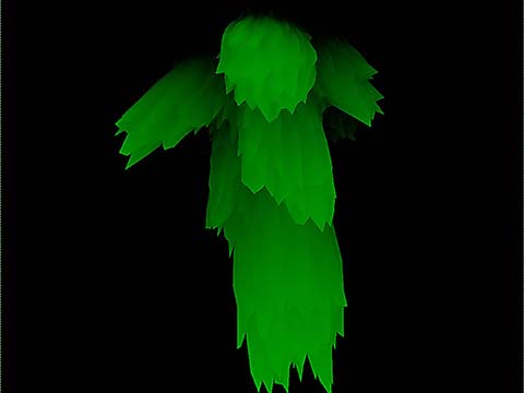
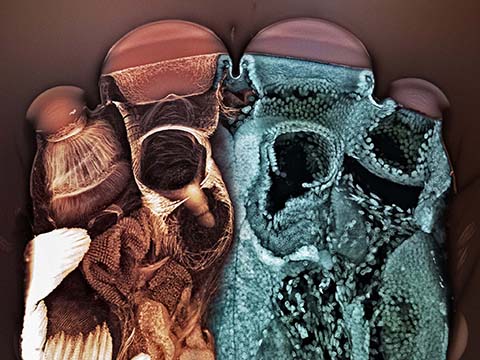
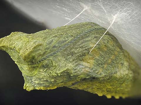
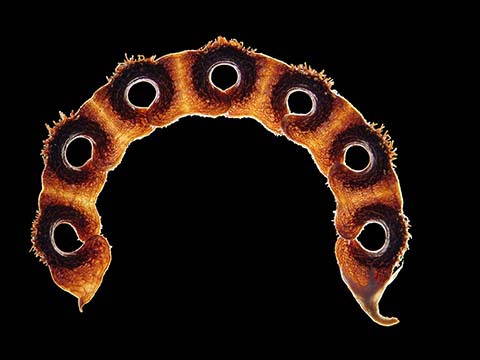
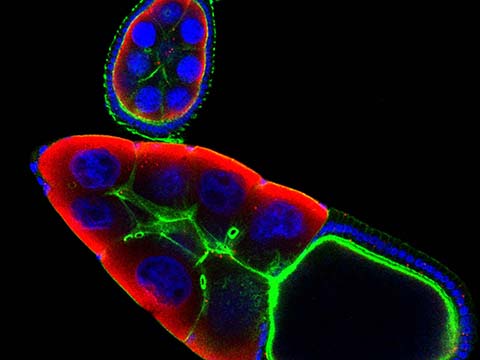
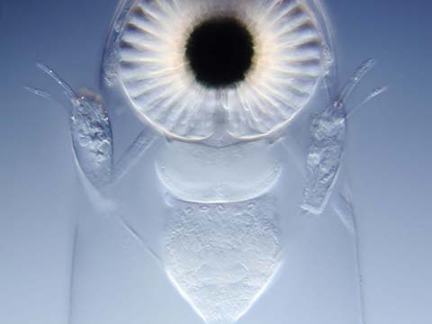
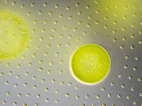
Honorable Mentions
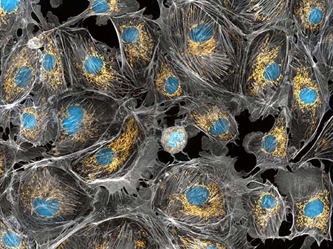
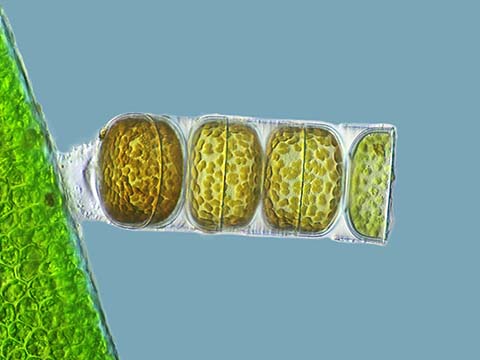
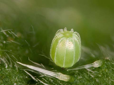
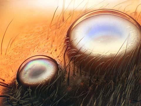
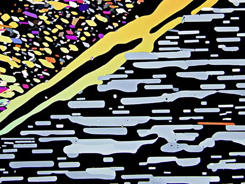
Images of Distinction

Judges
Alan Boyle
Science Editor
MSNBC.com
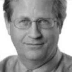
As MSNBC.com‘s science editor, Alan Boyle runs a virtual curiosity shop of the physical sciences and space exploration, plus paleontology, archaeology and other ologies that strike his fancy. Since joining MSNBC.com in 1996, Boyle has won awards from the National Academies, the American Association for the Advancement of Science, the National Association of Science Writers, the Society of Professional Journalists, the Space Frontier Foundation, the Pirelli Relativity Challenge and the CMU Cybersecurity Journalism Awards program.
He is the author of “The Case for Pluto,” a contributor to “A Field Guide for Science Writers,” the blogger behind Cosmic Log — and an occasional talking head on the MSNBC cable channel. During his 34 years of daily journalism in Cincinnati, Spokane and Seattle, he’s survived a hurricane, a volcanic eruption, a total solar eclipse and an earthquake. He has faith he’ll survive the Internet as well.
Richard Day, Ph.D.
Professor, Department of Cellular and Integrative Physiology
Indiana University School of Medicine
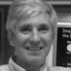 Richard Day is a Professor in the Department of Cellular and Integrative Physiology at the Indiana University School of Medicine. His research focuses on understanding the network of regulatory protein interactions that function to control cell-type specific gene expression. His laboratory group uses biochemical and molecular approaches to define networks of protein interactions that are coordinated by specific transcription factors. These in vitro approaches are then complemented by non-invasive live-cell imaging techniques using the many different color variants of the marine invertebrate fluorescent proteins. Recent studies from the laboratory have used Förster resonance energy transfer (FRET)-based microscopy approaches to begin to define networks of protein interactions in living cells.
Dan Vergano
Science Columnist
USA Today
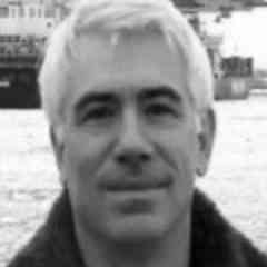
Dan Vergano covers science and society at USA TODAY, where he has worked since 1999. Previous reporting stints include Medical Tribune, HealthWeek (PBS) and Science News, as well as freelance reporting for Science, New Scientist, Men’s Health, The Washington Post and others. He was a 2008 Nieman Fellow for Journalism at Harvard, and most recently was the winner of the 2011 Gene Stuart science-writing award from the Society for American Archeology. Prior to reporting, Dan worked as a policy analyst and engineer for a federally-funded research and development center.
Simon C. Watkins Ph.D.
Professor and Vice Chairman for Department of Cell Biology and Physiology at the University Of Pittsburgh School of Medicine Founder and Director
University of Pittsburg Center for Biological Imaging
Dr. Watkins is a Professor and Vice Chairman for Department of Cell Biology and Physiology at the University Of Pittsburgh School Of Medicine. He is also the Founder and Director of the Center for Biologic Imaging. His current research focus is understanding the mechanisms of communication between cells of the immune system using molecular and optical imaging tools. To date, he has produced approximately 500 peer reviewed papers.
Dr. Watkins is actively involved in medical student and graduate student education, directing the graduate program in Cell Biology and Molecular Physiology for 10 years and serving on multiple current graduate student committees. He is also actively involved in medical education and was awarded an excellence in teaching award in 2005. Dr. Watkins is a Fellow of the Royal College of Pathologists, was appointed as the Raine Professor at the University of Western Australia in Perth in 1999 and 2008 and holds an honorary doctorate from the University of Umea in Sweden for his contributions to the field of microscopic imaging.
Judge’s Consultant
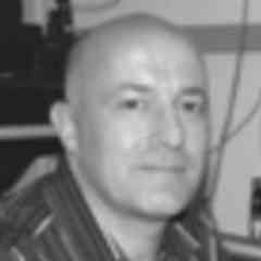
Michael W. Davidson
Director, Optical and Magneto-Optical Imaging Center at the National High Magnetic Field Laboratory
Florida State University
 Michael Davidson is the director of the Optical and Magneto-Optical Imaging Center at the National High Magnetic Field Laboratory at Florida State University. Involved with various aspects of microscopy for over 25 years, Davidson's scientific interests include the packaging of DNA into virus heads, liquid crystallinity in biological systems and the adsorption of small liquid crystal molecules onto surfaces. Davidson has authored many scientific articles on the subject of photomicrography and his photomicrographs have been published in more than a thousand national and international scientific journals, popular magazines and newspapers. In addition, Davidson's photomicrography has won more than 40 awards in scientific and industrial photography competitions and has been exhibited at over 50 locations nationwide. He is also the expert behind the Nikon Instruments educational Web site MicroscopyU (which can be access through the Nikon Instruments Web site at www.nikoninstruments.com) and his own www.molecularexpressions.com.