Digital Image and Video Galleries
Recording of images observed in the optical microscope is of paramount importance for students, teachers, scientists, and technicians in both educational and industrial environments. The MicroscopyU digital image galleries feature over 1,500 still images and videos that present a wide spectrum of specimens imaged using the classical illumination techniques of brightfield, differential interference contrast (DIC), fluorescence, Rheinberg, darkfield, reflected light, Hoffman modulation contrast, phase contrast, and polarized light.
Technique Galleries
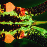
Confocal Microscopy
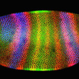
Enjoy the beauty of autofluorescence in thick sections of animal and plant tissues.
Movies
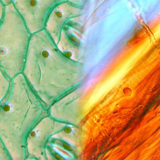
Differential Interference Contrast (DIC)
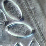
Compare specimen contrast using these complementary imaging techniques.
Image Comparisons • Movies
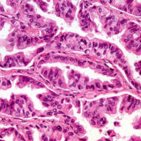
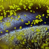
Fluorescence Microscopy
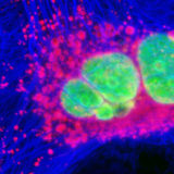
Cells and tissues examined with synthetic fluorophores in fluorescence microscopy.
Images
Phase Contrast
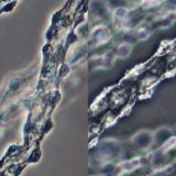
Specimen viewfields examined using positive and negative phase contrast.
Image Comparisons
Polarized Light
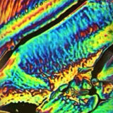
Time-lapse videos of chemicals as they change from solid to the liquid state.
Image Comparisons
Application Galleries
Digital Eclipse Image Gallery
Explore the performance of Nikon's DXM-1200 camera using several contrast techniques.
Images
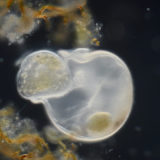
Nikon’s Small World
Nikon’s Small World is regarded as the leading forum for showcasing the beauty and complexity of life as seen through the light microscope. For over forty years, the Nikon Small World Competition has brought together photomicrographers of all levels and disciplines, and united them in a competition that truly celebrates and honors the intersection of science with art.
Photomicrography Competition
Nikon's Small World photomicrography competition recognizes excellence in photography through the light microscope.
Images
Small World in Motion Competition
The video competition encompasses any movie or digital time-lapse photography taken through the microscope.
Movies
Share this page:


