Polarized Light Microscopy
Polarized light is a contrast-enhancing technique that improves the quality of the image obtained with birefringent materials when compared to other techniques such as darkfield and brightfield, differential interference contrast, phase contrast, Hoffman modulation contrast, and fluorescence. Polarized light microscopes have a high degree of sensitivity and can be utilized for both quantitative and qualitative studies targeted at a wide range of anisotropic specimens. Qualitative polarizing microscopy is very popular in practice, with numerous volumes dedicated to the subject. In contrast, the quantitative aspects of polarized light microscopy, which is primarily employed in crystallography, represent a far more difficult subject that is usually restricted to geologists, mineralogists, and chemists. However, steady advances made over the past few years have enabled biologists to study the birefringent character of many anisotropic sub-cellular assemblies.
Review Articles
Introduction to Polarized Light (日本語)
Discussion of birefringence, Brewster's angle, and various forms of polarized light.
Principles of Birefringence (日本語)
Defined as double refraction of light in a transparent, molecularly ordered material.
Polarized Light Microscopy (日本語)
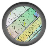
Using crossed polarized illumination to examine birefringent materials.
Principles of Interference
The addition or subtraction of light waves to produce constructive or destructive results.
Interactive Tutorials
-

Birefringent Crystals in Polarized Light
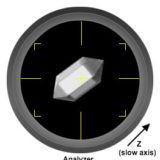
Explore how birefringent anisotropic crystals interact with polarized light in an optical microscope as the circular stage is rotated through 360 degrees.
-

Polarizer Rotation and Specimen Birefringence
Discover how specimen birefringence is affected by the angle of polarizer when observed in a polarized light microscope.
Related Nikon Products
Selected Literature References
Polarized Light Microscopy
Observing birefringent specimens using cross-polarized illumination.
Specimen Contrast in Microscopy
Examine the origins of contrast in a wide spectrum of specimens.
Contributing Authors
Philip C. Robinson - Department of Ceramic Technology, Staffordshire Polytechnic, College Road, Stroke-on-Trent, ST4 2DE United Kingdom.
Douglas B. Murphy - Department of Cell Biology and Microscope Facility, Johns Hopkins University School of Medicine, 725 N. Wolfe Street, 107 WBSB, Baltimore, Maryland 21205.
Kenneth R. Spring - Scientific Consultant, Lusby, Maryland, 20657.
Thomas J. Fellers, Matthew J. Parry-Hill, Brian O. Flynn, and Michael W. Davidson - National High Magnetic Field Laboratory, 1800 East Paul Dirac Dr., The Florida State University, Tallahassee, Florida, 32310.


