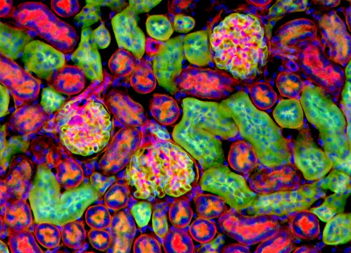
Mouse Kidney Tissue
Alexa Fluor 488 conjugated to the lectin wheat germ agglutinin, which selectively binds to N-acetylglucosamine and N-acetylneuraminic (sialic acid) residues and is frequently used to localize the Golgi network in mammalian cells, was utilized to label the mouse kidney tissue sample illustrated in the digital image above. The specimen was labeled for filamentous actin with Alexa Fluor 568 conjugated to phalloidin and for nuclear DNA with DAPI. Images were recorded in grayscale with a 12-bit digital camera coupled to either a Nikon E-600 or Eclipse 80i microscope equipped with bandpass emission fluorescence filter optical blocks. During the processing stage, individual image channels were pseudocolored with RGB values corresponding to each of the fluorophore emission spectral profiles.
Featured in:
Share this page:


