Digital Imaging in Optical Microscopy
This section explores new concepts in digital imaging technology and reviews both fundamental concepts and advanced techniques involved in digital imaging. In addition, several of the current camera systems designed for optical microscopy are explored.
Review Articles
Color Balance in Digital Imaging
The acquisition of color balanced images in the optical microscope can be a challenge.
Digital Imaging – New Opportunities for Microscopy
Digital imaging is applied to image capture for microscopy for high resolution and color fidelity in limited light conditions.
Digital Networking Camera Technology
Peter Drent explains how digital imaging has given microscopists a new medium.
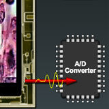
Fundamentals of Digital Imaging
Improvements in electronic camera and computer technology have made digital imaging cheaper and easier to use than conventional photography.
Introduction to Charge-Coupled Devices (CCDs)
Fundamental properties of CCDs, including pixels, readout, noise, and timing.
Interactive Tutorials
-
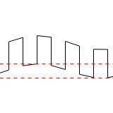
CCD Noise Sources
A review of the types of noise sources associated with digital imaging.
-
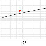
CCD Signal-To-Noise Ratio
Signal-to-noise represents the ratio of the measured light signal to the combined noise.
-
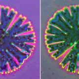
Color Balance in Digital Imaging
White and black balance settings on a digital camera can be used to adjust color balance.
-
Full-Frame CCD Operation
The readout scheme of a full-frame charge coupled device (CCD).
-
Matching Camera to Microscope Resolution
Vary numerical aperture, magnification, and video coupler size to match camera resolution.
-
Proximity-Focused Image Intensifiers
Light amplification with a micro-channel plate and photocathode.
-
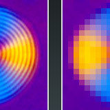
Spatial Resolution in Digital Imaging
Spatial resolution refers to the number of pixels utilized in construction of the image.
Related Nikon Products
Contributing Authors
Kenneth R. Spring - Scientific Consultant, Lusby, Maryland, 20657.
Peter Drent - Production Manager, Microscopy Division, Nikon Europe, Schipholweg 321, 1171PL Badhoevedorp, The Netherlands.
Matthew Parry-Hill, Thomas J. Fellers, and Michael W. Davidson - National High Magnetic Field Laboratory, 1800 East Paul Dirac Dr., The Florida State University, Tallahassee, Florida, 32310.
Share this page:


