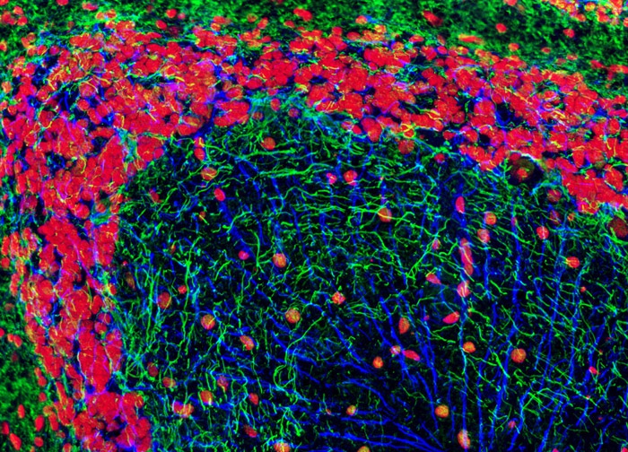
Brain Tissue Sample Labeled for GFAP, NF-P, and DNA
In order to visualize neurons and astroglia in a rat brain horizontal tissue section (shown above), the specimen was immunofluorescently labeled with mouse anti-NF-P and rabbit anti-GFAP primary antibodies followed by goat anti-mouse and anti-rabbit secondary antibodies conjugated to Alexa Fluor 568 (pseudocolored blue) and Alexa Fluor 488, respectively. Hoechst 33342 (pseudocolored red) was employed to counterstain cell nuclei. Images were recorded in grayscale with a 12-bit digital camera coupled to a Nikon Eclipse 80i microscope equipped with bandpass emission fluorescence filter optical blocks. During the processing stage, individual image channels were pseudocolored with RGB values corresponding to each of the fluorophore emission spectral profiles unless otherwise noted above.
Featured in:
Share this page:


