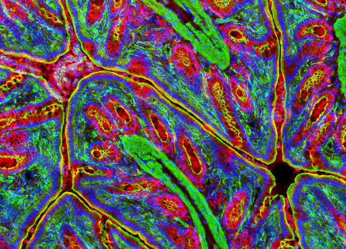
Golgi Networks and F-Actin Visualized in a Rat Colon Sample
In order to visualize lectin binding to the Golgi complex in a sample of rat colon, the tissue section above was treated with wheat germ agglutinin conjugated to Texas Red. The cells were subsequently counterstained with Alexa Fluor 488 conjugated to phalloidin to localize the filamentous actin network, and the nucleic acid stain Hoechst 33332 to label DNA in the nucleus. Images were recorded in grayscale with a 12-bit digital camera coupled to a Nikon Eclipse 80i microscope equipped with bandpass emission fluorescence filter optical blocks. During the processing stage, individual image channels were pseudocolored with RGB values corresponding to each of the fluorophore emission spectral profiles.
Featured in:
Share this page:


