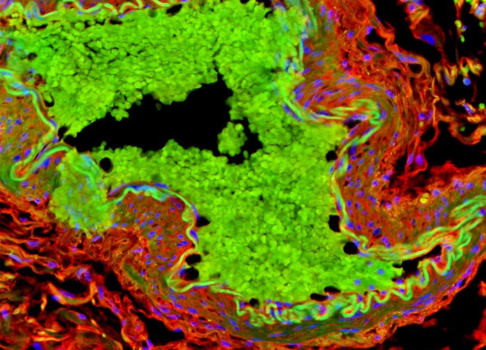
Human Small Intestine Tissue
The sample of human small intestine tissue presented in the digital image above was stained with Texas Red-X conjugated to wheat germ agglutinin (WGA), one of the most commonly used lectins in microbiology. WGA selectively binds to N-acetylglucosamine and N-acetylneuraminic (sialic acid) residues. In addition, the specimen was labeled with Alexa Fluor 488 conjugated to phalloidin, a phallotoxin that targets filamentous actin. DAPI, which emits blue fluorescence when it binds to AT regions of DNA, was employed as a counterstain. Images were recorded in grayscale with a 12-bit digital camera coupled to either a Nikon E-600 or Eclipse 80i microscope equipped with bandpass emission fluorescence filter optical blocks. During the processing stage, individual image channels were pseudocolored with RGB values corresponding to each of the fluorophore emission spectral profiles.
Featured in:
Share this page:


