Labeling Epididymis Tissue with Fluorescent Probes Conjugated to Phallotoxins and Lectins
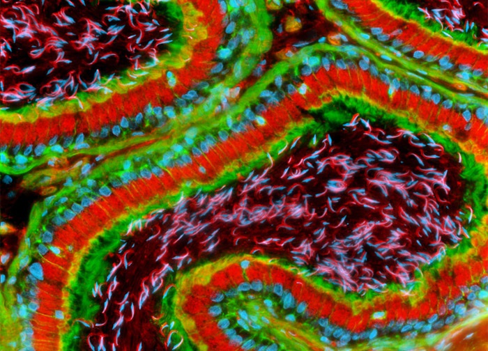
The rat epididymis tissue section illustrated in the digital image above was stained with Texas Red conjugated to wheat germ agglutinin, a plant-derived lectin that targets the Golgi apparatus, as well as Alexa Fluor 488 conjugated to phalloidin for cytoskeletal actin. Nuclei were labeled with Hoechst 33342. Images were recorded in grayscale with a 12-bit digital camera coupled to a Nikon Eclipse 80i microscope equipped with bandpass emission fluorescence filter optical blocks. During the processing stage, individual image channels were pseudocolored with RGB values corresponding to each of the fluorophore emission spectral profiles.
Featured in:
Share this page:


