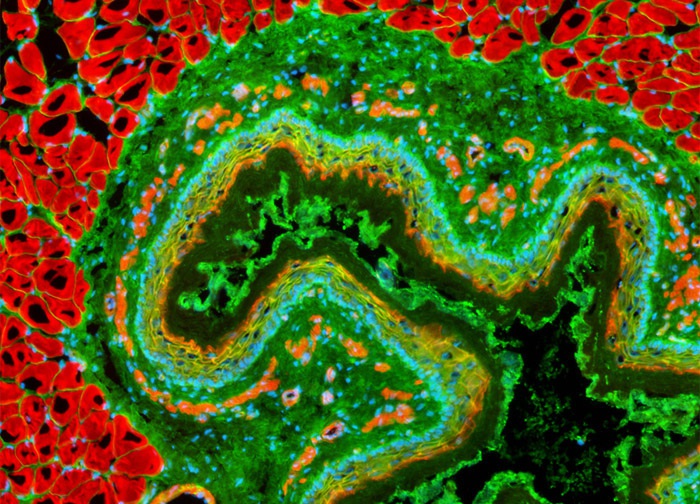
Lectin Carbohydrates and Filamentous Actin Visualized in a Rat Colon Sample
The rat esophagus tissue section illustrated in the digital image above was labeled with wheat germ agglutinin (WGA) conjugated to Oregon Green 488. WGA, which selectively binds to N-acetylglucosamine and N-acetylneuraminic (sialic acid) residues, is well suited for staining the Golgi network in fixed cells and tissues since a number of proteins and lipids found in the Golgi apparatus are glycosylated. The specimen was also labeled with Alexa Fluor 568 conjugated to phalloidin (targeting filamentous actin) and Hoechst 33342 (targeting DNA in the nucleus). Images were recorded in grayscale with a 12-bit digital camera coupled to a Nikon Eclipse 80i microscope equipped with bandpass emission fluorescence filter optical blocks. During the processing stage, individual image channels were pseudocolored with RGB values corresponding to each of the fluorophore emission spectral profiles.
Featured in:
Share this page:


