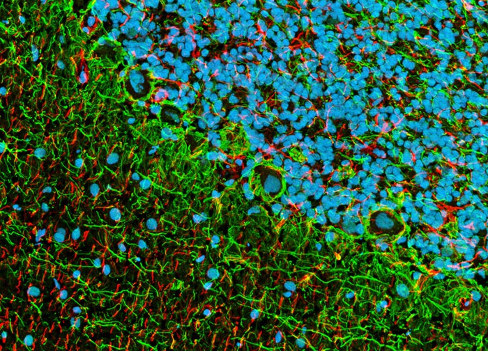
Localizing Fluorescent Tags to Neurofilaments and Astroglia in Brain Samples
In a double immunofluorescence experiment, this sagittal section of rat brain was fixed, permeabilized, blocked with 10-percent normal goat serum, and then treated with primary mouse antibodies against NF-P (phosphorylated neurofilaments) and rabbit antibodies against GFAP (glial fibrillary acidic protein). The primary targets were then visualized with goat anti-mouse secondary antibodies conjugated to Alexa Fluor 488 and anti-rabbit antibodies conjugated to Alexa Fluor 568. Hoechst 33342 was employed as a nuclear counterstain. Images were recorded in grayscale with a 12-bit digital camera coupled to a Nikon Eclipse 80i microscope equipped with bandpass emission fluorescence filter optical blocks. During the processing stage, individual image channels were pseudocolored with RGB values corresponding to each of the fluorophore emission spectral profiles.
Featured in:
Share this page:


