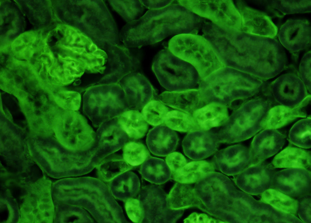
Mouse Kidney Tissue
BV-1A Longpass Emission (Narrow Bandwidth Excitation) Blue-Violet Set
Fluorescence emission intensity from a thin section of mouse kidney stained with multiple (3) fluorophores. Nuclei in the tissue section were targeted with the nucleic acid probe DAPI, which has an excitation maximum at 358 nanometers and an emission maximum at 461 nanometers when bound to DNA in cell cultures and tissue sections. In addition, the cryostat section was also simultaneously stained with Alexa Fluor 488 wheat germ agglutinin (glomeruli and convoluted tubules) and Alexa Fluor 568 phalloidin (filamentous actin and the brush border). Note the absence of signal from the red (Alexa Fluor 568) fluorophore, but the significant amount of fluorescence from the green (Alexa Fluor 488) probe. With an ultraviolet excitation longpass emission filter set, the darker nuclei in the specimen, which are devoid of cyan fluorescence emission intensity in the image above, would appear bright blue.


