Mouse Brain Tissue Sections
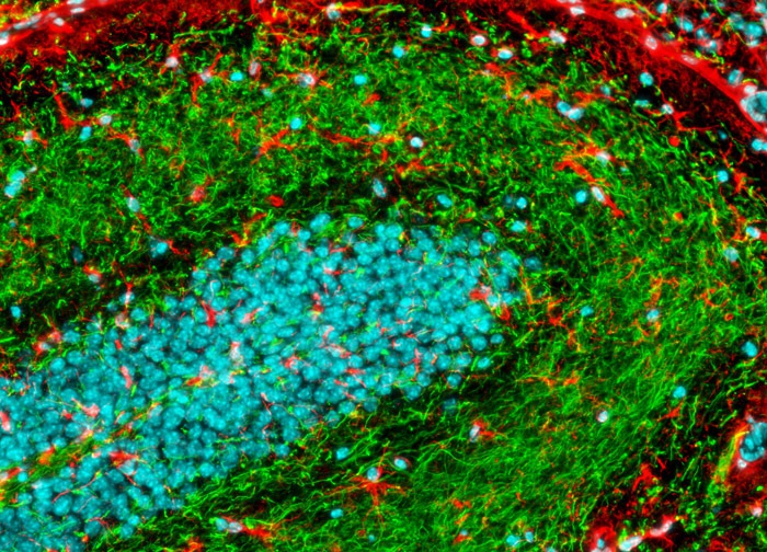
The neuron-specific intermediate filaments known as neurofilaments were targeted in the mouse brain coronal thin section shown above with chicken anti NF-H antibodies followed by goat anti-chicken secondary antibodies conjugated to Alexa Fluor 488. In addition, glial fibrillary acidic protein, an intermediate filament protein that is a key structural element of astroglia, was immunofluorescently labeled with primary anti-GFAP rabbit monoclonal antibodies followed by goat anti-rabbit Fab fragments conjugated to Alexa Fluor 568. Hoechst 33342 was employed as a nuclear counterstain. Images were recorded in grayscale with a 12-bit digital camera coupled to a Nikon Eclipse 80i microscope equipped with bandpass emission fluorescence filter optical blocks. During the processing stage, individual image channels were pseudocolored with RGB values corresponding to each of the fluorophore emission spectral profiles.
Featured in:
Share this page:


