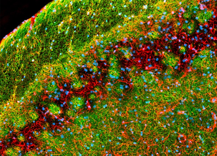
Neural Tissue Labeled for GFAP, Myelin BP, and DNA
Myelin basic protein, which is a marker for the fatty sheaths surrounding the axons of myelinated nerve fibers, and glial fibrillary acidic protein, a type III intermediate filament protein found primarily in astroglia, were immunofluorescently labeled in the rat brain sagittal tissue section presented above by treating the specimen with a cocktail of mouse anti-myelin BP and rabbit anti-GFAP primary antibodies followed by goat anti-mouse and anti-rabbit secondary antibodies conjugated to Alexa Fluor 488 and Alexa Fluor 568, respectively. Hoechst 33342, a dsDNA-interactive agent, was utilized to target cell nuclei. Images were recorded in grayscale with a 12-bit digital camera coupled to a Nikon Eclipse 80i microscope equipped with bandpass emission fluorescence filter optical blocks. During the processing stage, individual image channels were pseudocolored with RGB values corresponding to each of the fluorophore emission spectral profiles.
Featured in:
Share this page:


