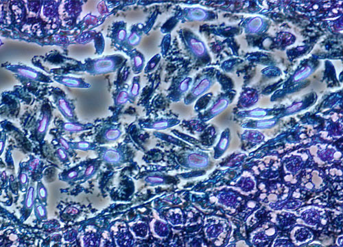
Nucleic Acid Stains in Animal Cells

Cells are the basic building blocks of all living organisms. While describing plant tissues in 1665, Robert Hooke first coined the term because the cellulose walls of cork that he was able to see through his microscope reminded him of the cells inhabited by monks. The images presented below compare a thin section of animal tissue stained for the nucleic acids RNA and DNA.
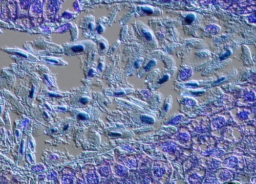
DIC
Phase

Phase

The finer points of cellular purpose and composition were not understood form many years, slowly developing as microscopes improved. In fact, the realization that cells were the fundamental unit of plants and animals did not occur until the 1830s. Previously cells had been believed to be simply pores, but the detection of nuclei and moving protoplasm put an end to the misconception. For other discoveries, additional microscopical enhancements needed to be made. Walter Flemming's advanced methods of fixing and staining cells, for instance, facilitated his observation of chromosome transmission between cells and led to his very precise account of mitosis and cell division in 1882.

DIC

Recent experimentation has been aimed at utilizing animal cells for human recovery from diseases, such as Parkinson's. Scientists hope to use cloned cells to replace improperly functioning cells in patients, thereby reducing tremors and other symptoms. Although the research is in its early stages, if it proves successful the medical world could be revolutionized. However, concerns such as the possibility of transmitting animal disease to humans through cell transplantation and the ethics of genetic cloning are problems that may hinder future developments.

Share this page: