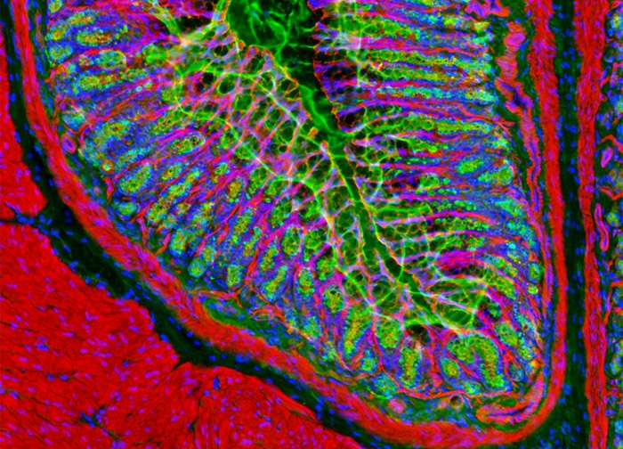
Probing Golgi Networks and Filamentous Actin Networks in a Sample of Rat Stomach Tissue
In the digital image above, a rat stomach tissue section excised from the pylorus region is presented that was labeled with the fluorophore Oregon Green 488 conjugated to wheat germ agglutinin, targeting the Golgi network. The sample was also labeled with Alexa Fluor 568 conjugated to phalloidin and Hoechst 33342, which target filamentous actin and nuclear DNA, respectively. Images were recorded in grayscale with a 12-bit digital camera coupled to a Nikon Eclipse 80i microscope equipped with bandpass emission fluorescence filter optical blocks. During the processing stage, individual image channels were pseudocolored with RGB values corresponding to each of the fluorophore emission spectral profiles.
Featured in:
Share this page:


