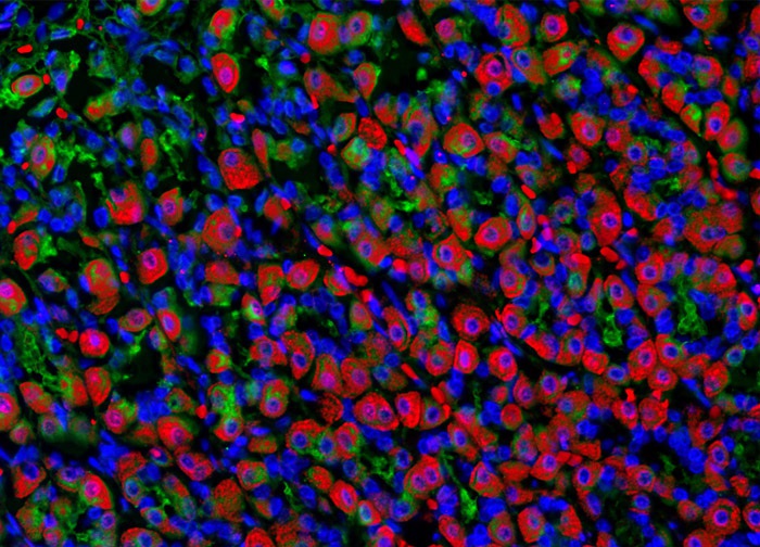
Probing Rat Tissue Sections with Alexa Fluor 488 Conjugated to a Lectin Isolated from the Red Kidney Bean
Alexa Fluor 488 conjugated to a lectin isolated from the red kidney bean (Phaseolus vulgaris) was utilized to target glycoproteins present in the sample of rat stomach tissue depicted in the digital image above (yielding green emission). The specimen was also stained for F-actin and nuclear DNA with Alexa Fluor 568 (red emission) conjugated to phalloidin and Hoechst 33258 (blue emission), respectively. Images were recorded in grayscale with a 12-bit digital camera coupled to either a Nikon E-600 or Eclipse 80i microscope equipped with bandpass emission fluorescence filter optical blocks. During the processing stage, individual image channels were pseudocolored with RGB values corresponding to each of the fluorophore emission spectral profiles.
Featured in:
Share this page:


