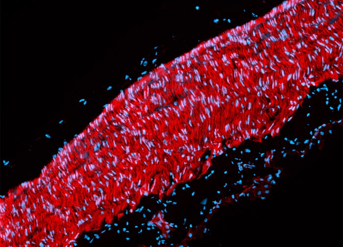
Rat Aorta Tissue Section
The rat aorta tissue section presented in the digital image above was labeled for the filamentous actin cytoskeletal network with Alexa Fluor 568 conjugated to phalloidin, a cyclic peptide produced by the Amanita phalloides mushroom. Cell nuclei were counterstained with Hoechst 33342. Images were recorded in grayscale with a 12-bit digital camera coupled to a Nikon Eclipse 80i microscope equipped with bandpass emission fluorescence filter optical blocks. During the processing stage, individual image channels were pseudocolored with RGB values corresponding to each of the fluorophore emission spectral profiles.
Featured in:
Share this page:


