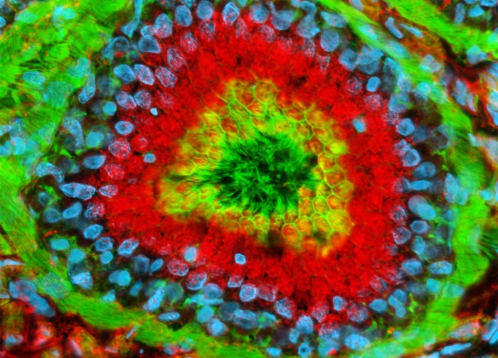
Rat Epididymis Tissue Section
In the digital image above, a rat epididymis tissue section is presented that was labeled with the fluorophore Texas Red conjugated to wheat germ agglutinin, a fluorescent lectin that selectively binds to sialic acid residues. Wheat germ agglutinin conjugates are often used as probes for the Golgi network in mammalian tissues and cells. The sample was also stained with Alexa Fluor 488 conjugated to phalloidin and Hoechst 33342, which target filamentous actin and nuclear DNA, respectively. Images were recorded in grayscale with a 12-bit digital camera coupled to a Nikon Eclipse 80i microscope equipped with bandpass emission fluorescence filter optical blocks. During the processing stage, individual image channels were pseudocolored with RGB values corresponding to each of the fluorophore emission spectral profiles.
Featured in:
Share this page:


