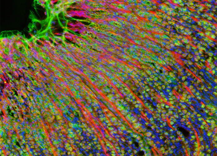
Rat Stomach Fundus Region Tissue Section Labeled with Fluorescent Probes Conjugated to Phallotoxins and Lectins
This widefield image of a rat stomach tissue section was produced by probing the specimen with Alexa Fluor 568, Oregon Green 488, and Hoechst 33342. The Alexa Fluor dye was conjugated to phalloidin, targeting the cytoskeletal filamentous actin network, and Oregon Green 488 was conjugated to WGA in order to label the Golgi complex. Cell nuclei were visualized with Hoechst 33342. Images were recorded in grayscale with a 12-bit digital camera coupled to a Nikon Eclipse 80i microscope equipped with bandpass emission fluorescence filter optical blocks. During the processing stage, individual image channels were pseudocolored with RGB values corresponding to each of the fluorophore emission spectral profiles.
Featured in:
Share this page:


