Rat Thoracic Aorta Cellular Mitochondria
BV-2A Longpass Emission (Medium Bandwidth Excitation) Blue-Violet Set
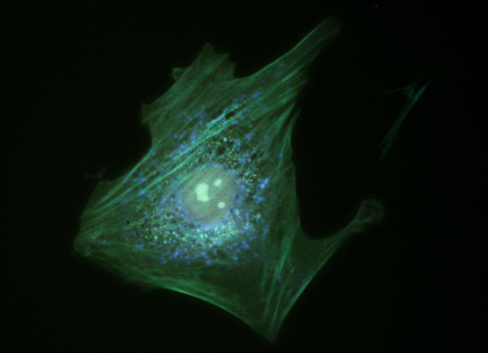
Fluorescence emission intensity from a culture of rat thoracic aorta (muscle) cells that were immunofluorescently labeled with primary anti-oxphos complex V inhibitor protein monoclonal antibodies (mouse) followed by goat anti-mouse Fab fragments conjugated to Pacific Blue. The absorption maximum of Pacific Blue is 410 nanometers and the emission maximum occurs at 455 nanometers. In addition, the specimen was simultaneously stained for F-actin with Alexa Fluor 488 (green) conjugated to phalloidin, and for DNA with SYTOX Orange. Note the presence of significant signal from the green fluorophore, which clearly delineates the cytoskeletal actin network. Blue mitochondria are also evident surrounding the nucleus and throughout the cytoplasm. Fluorescence emission from SYTOX Orange, which is primarily localized in the nucleus, appears green under these observation conditions. In many cases, SYTOX Orange stains a variety of cytoplasmic elements in addition to DNA.
Share this page:


