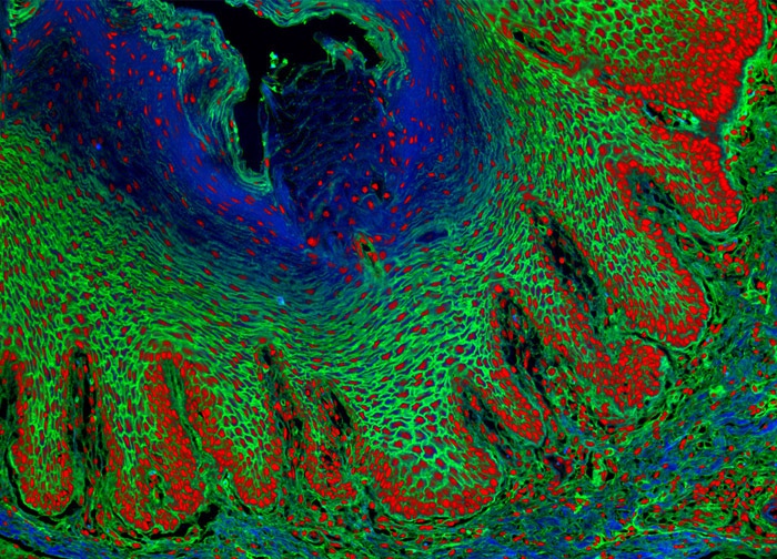
Sheep Tongue Tissue
In order to localize a blue fluorescent tag to F-actin in the sheep tongue tissue sample presented in the digital image above, the specimen was labeled with Alexa Fluor 350 conjugated to phalloidin, a commonly used actin-binding phallotoxin. Oregon Green 488 conjugated to the lectin wheat germ agglutinin, which selectively binds to N-acetylglucosamine and N-acetylneuraminic residues, was also applied to the tissue sample. Cell nuclei were counterstained with SYTOX Orange. Images were recorded in grayscale with a 12-bit digital camera coupled to either a Nikon E-600 or Eclipse 80i microscope equipped with bandpass emission fluorescence filter optical blocks. During the processing stage, individual image channels were pseudocolored with RGB values corresponding to each of the fluorophore emission spectral profiles.
Featured in:
Share this page:


