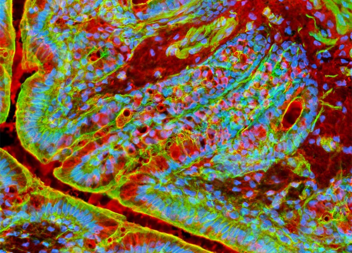
Sialic Acid Residues, Filamentous Actin, and Nuclear DNA in a Rat Rectum Tissue Sample
In order to localize a green fluorescent tag to F-actin in the sample of rat rectum tissue presented in the digital image above, the specimen was labeled with Alexa Fluor 488 conjugated to phalloidin, a cyclic peptide derived from the toxic death cap fungus (Amanita phalloides). Texas Red conjugated to the lectin wheat germ agglutinin, which selectively binds to N-acetylglucosamine and N-acetylneuraminic residues, was also applied to the tissue sample, as was the nuclear counterstain Hoechst 33342. Images were recorded in grayscale with a 12-bit digital camera coupled to a Nikon Eclipse 80i microscope equipped with bandpass emission fluorescence filter optical blocks. During the processing stage, individual image channels were pseudocolored with RGB values corresponding to each of the fluorophore emission spectral profiles.
Featured in:
Share this page:


