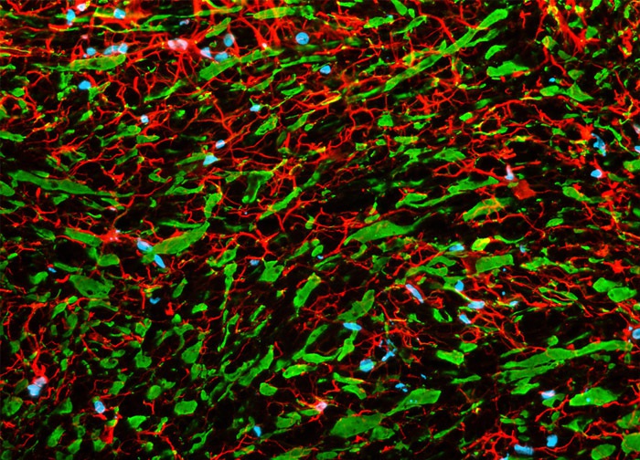
Utilizing Antibodies to Target Neurofilaments and Astrocytes
This widefield fluorescence image of a rat brain tissue section was produced by probing the specimen with Alexa Fluor 488, Alexa Fluor 568, and Hoechst 33342. The two Alexa Fluor dyes were conjugated to secondary antibodies directed against primary mouse anti-NF-P antibodies and rabbit anti-GFAP antibodies in order to label phosphorylated neurofilaments expressed in neurons (Alexa Fluor 488) and glial fibrillary acidic protein in astrocytes and certain other astroglia (Alexa Fluor 568). The nuclear counterstain Hoechst 33342 was employed to visualize cell nuclei. Images were recorded in grayscale with a 12-bit digital camera coupled to a Nikon Eclipse 80i microscope equipped with bandpass emission fluorescence filter optical blocks. During the processing stage, individual image channels were pseudocolored with RGB values corresponding to each of the fluorophore emission spectral profiles.
Featured in:
Share this page:


