Enhanced Yellow Fluorescent Protein (EYFP) Actin Filament Localization
The network of fibrous actin filaments (also commonly referred to as microfilaments) found in the vast majority of mammalian cell lines appears as a complex interconnected organization of linear bundles aligned in two-dimensional arrays or as a three-dimensional matrix. These protein fibers are easily labeled with synthetic or natural fluorophores and can be subsequently visualized using fluorescence microscopy, as illustrated for a variety of adherent cell lines in Figure 1. The single bandpass emission filter featured by the Nikon YFP HYQ optical block, which was employed to capture these images, produces sharp contrast with little interference from autofluorescence or other fluorescent species.
Figure 1 - EYFP-Actin Subcellular Localization in Transfected Cells
Plasmid pEYFP-Actin vector gene product expression in various cell types (from both transiently and stably transfected clones; see Figure 1) occurs due to the efficient intracellular translation of a fusion nucleotide sequence combining the enhanced yellow fluorescent protein domain with a functional copy of human cytoplasmic beta-actin. When expressed in living cells, the gene product is incorporated into actin filaments without significant structural interference from the EYFP protein moiety. The fluorescence excitation maximum of EYFP is 513 nanometers and the corresponding emission maximum occurs at 527 nanometers, with a relatively high (approximately 0.60) fluorescence quantum yield. In addition to the four chromophore mutations that shift the fluorescence emission maximum, the nucleotide coding sequence of the EYFP gene contains over 190 silent base alterations, which correspond to human codon-usage preferences that increase translational efficiency.
The collection of specimens illustrated in Figure 1 demonstrates the effectiveness of the Nikon YFP HYQ filter combination for imaging a series of cell lines containing the chimeric EYFP plasmid vector that expresses a fluorescent fusion protein targeted at the intracellular mitochondrial network. The fusion protein is transported into the mitochondria to enable visualization of the subcellular structure in living and fixed cells. Susceptible adherent cell cultures were transfected with the appropriate vector using proprietary lipophilic reagents, and were then cultured for a period of at least 24 hours in nutrient medium supplemented with fetal bovine serum to allow high expression levels of the fluorescent fusion protein.
The enhanced yellow fluorescent protein gene used in these studies contains four amino acid substitutions that shift the emission maximum of green fluorescent protein (GFP) by 18 nanometers, from approximately 509 to 527 nanometers. The gene is optimized with human codons (as described above) and features a consensus Kozak translation initiation signal to achieve higher expression levels in mammalian cell cultures. In general, vectors targeted at specific subcellular organelles contain a fusion gene segment, which couples the EYFP gene to a peptide sequence or complete protein that is localized to a region of interest in living cells.
Additional EYFP-Actin Images with the YFP HYQ Filter Combination
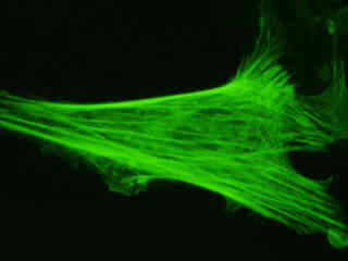
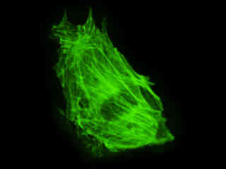
Rat Thoracic Aorta Myoblast Cell Actin
Fluorescence emission intensity from a culture of embryonic rat thoracic aorta medial layer (A-10 line) myoblast cells that were transfected with a pEYFP-Actin plasmid subcellular localization vector. Cells were transiently transfected and cultured in nutrient medium for a minimum of 24 hours before recording images.
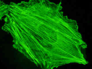
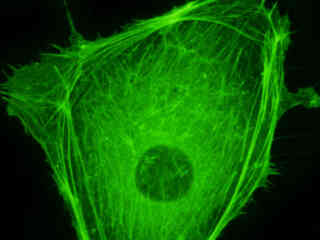
Rat Kangaroo Kidney Cell Actin
Fluorescence emission intensity from a culture of rat kangaroo kidney epithelial (PtK2 line) cells that were transfected with a pEYFP-Actin plasmid subcellular localization vector. Cells were transiently transfected and cultured in nutrient medium for a minimum of 24 hours before recording images.
Rat Kangaroo Kidney Cell Actin
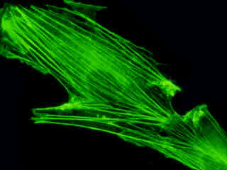
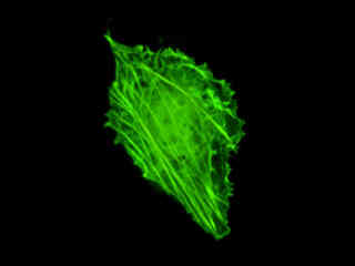
Indian Muntjac Deer Skin Cell Actin
Indian Muntjac Deer Skin Fibroblast Cell Actin
Observe the fluorescence emission in a culture of normal Indian Muntjac deer skin fibroblast cells that were transfected with a pEYFP-Actin plasmid subcellular localization vector. The enhanced yellow fluorescent protein gene used in these studies contains several important amino acid substitutions that shift the emission maximum of green fluorescent protein (GFP) by approximately 18 nanometers, from 509 to 527 nanometers.
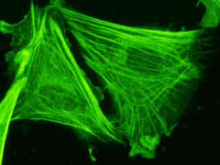
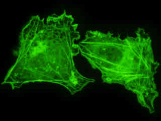
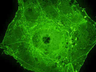
Human Bone Osteosarcoma Epithelial Cell Actin
Fluorescence emission intensity from a culture of human bone osteosarcoma (U2OS line) epithelial cells that were transfected with a pEYFP-Actin plasmid subcellular localization vector. Cells were transiently transfected and cultured in nutrient medium for a minimum of 24 hours before recording images.
Albino Swiss Mouse Embryonic Cell Actin
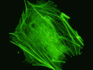
Examine the fluorescence emission intensity from a culture of albino Swiss mouse embryonic fibroblast (3T3 line) cells that were transfected with a pEYFP-Actin plasmid subcellular localization vector. Note the complex two and three-dimensional network of actin filaments and stress fibers that extend throughout the cytoplasm and inhabit the cortex in this cell line.
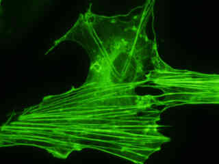
Bovine Pulmonary Artery Cell Actin
Fluorescence emission intensity from a culture of bovine pulmonary artery endothelial (BPAE line) cells that were transfected with a pEYFP-Actin plasmid subcellular localization vector. Cells were transiently transfected and cultured in nutrient medium for a minimum of 24 hours before recording images.
Normal African Green Monkey Kidney Cell Actin
Fluorescence emission intensity from a culture of normal African green monkey kidney fibroblast cells (CV-1 line) that were transfected with a pEYFP-Actin plasmid subcellular localization vector. Cells were transiently transfected and cultured in nutrient medium for a minimum of 24 hours before recording images.
Transformed African Green Monkey Kidney Cell Actin
Fluorescence emission intensity from a culture of transformed (SV-40) African green monkey kidney fibroblast cells (COS-7 line) that were transfected with a pEYFP-Actin plasmid subcellular localization vector. Cells were transiently transfected and cultured in nutrient medium for a minimum of 24 hours before recording images.
Contributing Authors
Related Nikon Products
Share this article:


