Enhanced Yellow Fluorescent Protein (EYFP) Endosome Localization
In eukaryotic cells, endosomes constitute a large network of cytoplasmic vesicles that are formed through the fusion of smaller internalized vesicles arising from receptor-mediated endocytosis. Often, the endosomal vesicles ultimately transfer their contents to the lysosomes for processing, but this is not always the case. Recombinant plasmids have been constructed that contain a fusion protein consisting of the yellow-green variant (referred to as enhanced yellow fluorescent protein; EYFP) of the Aequorea victoria green fluorescent protein (GFP) coupled to human RhoB GTPase, an enzyme that is localized in early endosomes, recycling endosomes, and multivesicular bodies. Upon transcription and translation of the plasmid in transfected mammalian hosts, the fused RhoB domain is responsible for transport and distribution of the fluorescent protein chimera throughout the cellular endosomal network.
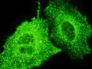
Figure 1 - EYFP-Endosomes Subcellular Localization in Transfected Cells
The human RhoB GTPase enzyme fused to an enhanced yellow fluorescent protein domain can be efficiently incorporated into the endosomes from a variety of mammalian cell lines (as illustrated in Figure 1). This chimera is entirely localized to both early and late endosomes and does not display a significant effect on endosomal trafficking. The RhoB GTPase-EYFP vector also contains a c-Myc epitope that enables identification of the fusion protein using immunofluorescence techniques in fixed cells. Intracellular endosomal networks labeled with resident macromolecules containing fluorescent protein domains, such as GFP or EYFP, can be readily visualized using fluorescence microscopy, as illustrated for a variety of established adherent cell lines in Figure 1. The single bandpass emission filter featured by the Nikon YFP HYQ optical block, which was employed to capture all of these images, produces sharp contrast with little interference from autofluorescence or other fluorescent species.
Plasmid pEYFP-Endosome vector gene product expression in various cell types (from both transiently and stably transfected clones; see Figure 1) occurs due to the efficient intracellular translation of a fusion nucleotide sequence combining the enhanced yellow fluorescent protein domain with human RhoB GTPase, as discussed above. Simian virus 40 (SV40) polyadenylation signals inserted downstream from the EYFP-Endosome fusion sequence direct proper processing of the 3' end of the transcribed messenger RNA. The fluorescence excitation maximum of EYFP is 513 nanometers and the corresponding emission maximum occurs at 527 nanometers, with a relatively high (approximately 0.60) fluorescence quantum yield. In addition to the four chromophore mutations that shift the fluorescence emission maximum, the nucleotide coding sequence of the EYFP gene contains over 190 silent base alterations, which correspond to human codon-usage preferences that are designed to increase translational efficiency.
The collection of specimens illustrated in Figure 1 demonstrates the effectiveness of the Nikon YFP HYQ filter combination for imaging a variety of cell lines that express gene products from the EYFP plasmid vector sequence for a fluorescent fusion protein chimera targeted at the intracellular endosomal network. Susceptible adherent cell cultures were transfected with the appropriate vector using proprietary lipophilic reagents, and were then cultured for a period of at least 24 hours in nutrient medium supplemented with fetal bovine serum to allow high expression levels of the fluorescent fusion protein.
The enhanced yellow fluorescent protein gene used in these studies contains four amino acid substitutions that shift the emission maximum of green fluorescent protein (GFP) by 18 nanometers, from approximately 509 to 527 nanometers. The gene is optimized with human codons (as described above) and features a consensus Kozak translation initiation signal to achieve higher expression levels in mammalian cell cultures. In general, vectors targeted at specific subcellular organelles contain a fusion gene segment, which couples the EYFP gene to a peptide sequence or complete protein that is localized to a region of interest in living cells.
Additional EYFP-Endosome Images with the YFP HYQ Filter Combination
HeLa Human Cervical Carcinoma Cellular Endosomes
Fluorescence emission intensity from a culture of human cervical adenocarcinoma (HeLa line) epithelial cells that were transfected with a pEYFP-Endosome plasmid subcellular localization vector.
Rat Thoracic Aorta Medial Layer Cellular Endosomes
Fluorescence emission intensity from a culture of rat thoracic aorta medial layer (A-10 line) myoblast cells that were transfected with a pEYFP-Endosome plasmid subcellular localization vector.
Rat Kangaroo Kidney Cellular Endosomes
Fluorescence emission intensity from a culture of rat kangaroo kidney (PtK2 line) epithelial cells that were transfected with a pEYFP-Endosome plasmid subcellular localization vector.
Canine Kidney Cellular Endosomes
Fluorescence emission intensity from a culture of canine kidney (Madin-Darby; MDCK line) epithelial cells that were transfected with a pEYFP-Endosome plasmid subcellular localization vector.
Human Osteosarcoma Cellular Endosomes
Fluorescence emission intensity from a culture of human fetal bone osteosarcoma (U2OSline) epithelial cells that were transfected with a pEYFP-Endosome plasmid subcellular localization vector.
Normal African Green Monkey Kidney Cellular Endosomes
Fluorescence emission intensity from a culture of normal African green monkey kidney (Vero line) epithelial cells that were transfected with a pEYFP-Endosome plasmid subcellular localization vector.
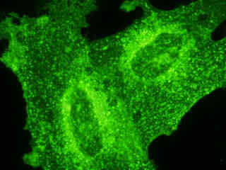
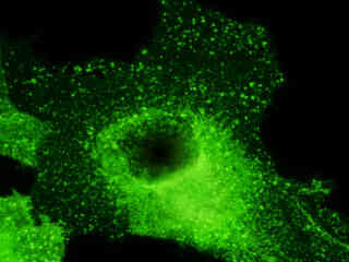
Rat Jejunum Myenteric Plexus Glial Cellular Endosomes
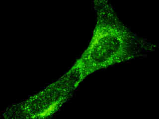
Examine the fluorescence emission intensity from a culture of rat glial (EGC line) cells that were transfected with a pEYFP-Endosome plasmid subcellular localization vector. Note the fluctuating level of green fluorescence defining the endosomal network, which tends to exhibit a higher degree of signal in areas adjacent to the nucleus.
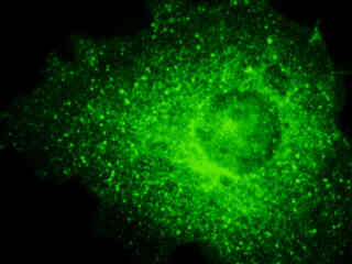
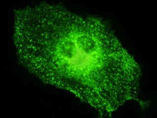
Mouse Endothelioma Cellular Endosomes
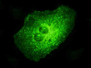
Mouse endothelial hemangioendothelioma (EOMA line) cells were transiently transfected and cultured in nutrient medium for a minimum of 24 hours before recording images. Note the generally high level of green fluorescence observed throughout the cytoplasm in this cell line.
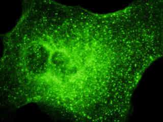
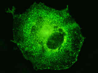
Normal African Green Monkey Kidney Cellular Endosomes
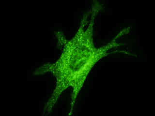
A culture of normal African green monkey kidney (CV-1 line) fibroblast cells was transfected with a pEYFP-Endosome plasmid subcellular localization vector, and then grown in nutrient medium for a minimum of 24 hours before recording images. Although the enhanced yellow fluorescent protein gene used in these studies contains several important amino acid substitutions that shift the emission maximum of green fluorescent protein, the fluorescence emission still appears green when observed in the microscope eyepieces.
Bovine Pulmonary Artery Endothelial Cellular Endosomes
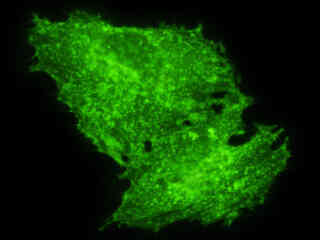
This section reviews fluorescence emission intensity observed with the Nikon YFP HYQ blue-green excitation filter combination in a culture of bovine pulmonary artery endothelial (BPAE) cells labeled with a chimera of enhanced yellow fluorescent protein (EYFP) and human RhoB GTPase, a protein that targets early and late endosomes.
Transformed African Green Monkey Kidney Endosomes
Review the fluorescence emission intensity from a culture of SV40-transformed African green monkey kidney (COS-7 line) fibroblast cells that were transfected with a pEYFP-Endosome plasmid subcellular localization vector. Note the fluctuating level of green fluorescence defining the endosomal network, which tends to exhibit a higher degree of signal in areas adjacent to the nucleus.
Contributing Authors
Related Nikon Products
Share this article:


