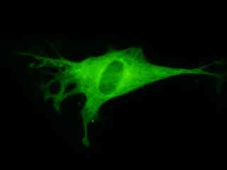
Enhanced Yellow Fluorescent Protein (EYFP) Tubulin Localization
Microtubules are cylindrical biopolymeric filaments that play an important structural and functional role in the dynamics of the cellular cytoskeleton. These hollow, but rigid, helical tubes are constructed with several variations of a protein known as tubulin. Among the many biological tasks assigned to microtubules is segregation of the chromosomes during mitosis, formation of cilia and flagella on the surface of a cell, and transport of materials through axons. Recombinant plasmids have been constructed that contain a fusion protein consisting of the yellow-green variant (referred to as enhanced yellow fluorescent protein; EYFP) of the Aequorea victoria green fluorescent protein (GFP) coupled to human alpha-tubulin, one of the tubulin subunits in microtubules. Upon transcription and translation of the plasmid in transfected mammalian hosts, the fused tubulin domain is responsible for transport and distribution of the fluorescent protein chimera throughout the cellular microtubule network.
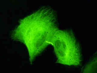
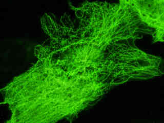
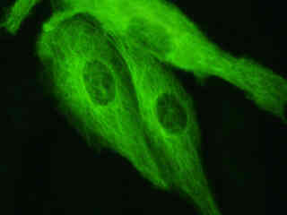
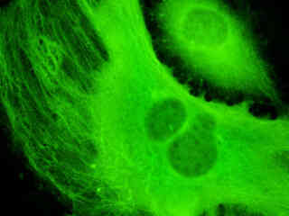
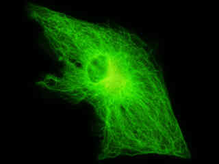
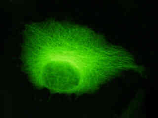
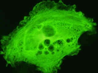
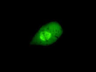
Figure 1 - EYFP-Tubulin Subcellular Localization in Transfected Cells
The human alpha-tubulin protein can be efficiently incorporated into the microtubules from a variety of mammalian cell lines (Figure 1). Because this monomer represents approximately 50 percent of the tubulin enlisted in the construction of microtubules, the resulting structures contain a significant level of fluorescent protein label. Intracellular microtubule networks labeled with fluorescent proteins, such as GFP or EYFP, can be readily visualized using fluorescence microscopy, as illustrated for a variety of established adherent cell lines in Figure 1. The single bandpass emission filter featured by the Nikon YFP HYQ optical block, which was employed to capture all of these images, produces sharp contrast with little interference from autofluorescence or other fluorescent species.
Plasmid pEYFP-Tubulin vector gene product expression in various cell types (from both transiently and stably transfected clones; see Figure 1) occurs due to the efficient intracellular translation of a fusion nucleotide sequence combining the enhanced yellow fluorescent protein domain with human alpha-tubulin, as discussed above. Simian virus 40 (SV40) polyadenylation signals inserted downstream from the EYFP-Tubulin fusion sequence direct proper processing of the 3' end of the transcribed messenger RNA. The fluorescence excitation maximum of EYFP is 513 nanometers and the corresponding emission maximum occurs at 527 nanometers, with a relatively high (approximately 0.60) fluorescence quantum yield. In addition to the four chromophore mutations that shift the fluorescence emission maximum, the nucleotide coding sequence of the EYFP gene contains over 190 silent base alterations, which correspond to human codon-usage preferences that are designed to increase translational efficiency.
The collection of specimens illustrated in Figure 1 demonstrates the effectiveness of the Nikon YFP HYQ filter combination for imaging a series of cell lines containing the chimeric EYFP plasmid vector that expresses a fluorescent fusion protein chimera targeted at the intracellular microtubule network. Susceptible adherent cell cultures were transfected with the appropriate vector using proprietary lipophilic reagents, and were then cultured for a period of at least 24 hours in nutrient medium supplemented with fetal bovine serum to allow high expression levels of the fluorescent fusion protein.
The enhanced yellow fluorescent protein gene used in these studies contains four amino acid substitutions that shift the emission maximum of green fluorescent protein (GFP) by 18 nanometers, from approximately 509 to 527 nanometers. The gene is optimized with human codons (as described above) and features a consensus Kozak translation initiation signal to achieve higher expression levels in mammalian cell cultures. In general, vectors targeted at specific subcellular organelles contain a fusion gene segment, which couples the EYFP gene to a peptide sequence or complete protein that is localized to a region of interest in living cells.
Additional EYFP-Tubulin Images with the YFP HYQ Filter Combination
Canine Kidney Cell Tubulin
Fluorescence emission intensity from a culture of normal canine kidney epithelial (Madin-Darby; MDCK line) cells that were transfected with a pEYFP-Tubulin plasmid subcellular localization vector.
Transformed African Green Monkey Kidney Cell Tubulin
Fluorescence emission intensity from a culture of simian virus 40 transformed African green monkey kidney (COS-7 line) fibroblast cells that were transfected with a pEYFP-Tubulin plasmid subcellular localization vector.
Bovine Pulmonary Artery Cell Tubulin
Fluorescence emission intensity from a culture of bovine pulmonary artery endothelial (BPAE line) cells that were transfected with a pEYFP-Tubulin plasmid subcellular localization vector.
Mouse Endothelioma Cell Tubulin
Fluorescence emission intensity from a culture of mouse endothelioma (EOMA line) cells that were transfected with a pEYFP-Tubulin plasmid subcellular localization vector.
Rat Kangaroo Kidney Cell Tubulin
Fluorescence emission intensity from a culture of rat kangaroo kidney epithelial (PtK2 line) cells that were transfected with a pEYFP-Tubulin plasmid subcellular localization vector.
HeLa Cell Tubulin
Fluorescence emission intensity from a culture of human cervical carcinoma epithelial (HeLa line) cells that were transfected with a pEYFP-Tubulin plasmid subcellular localization vector.
Rat Thoracic Aorta Myoblast Cell Tubulin
Examine the fluorescence emission intensity from a culture of embryonic rat thoracic aorta medial layer (A-10 line) myoblast cells that were transfected with a pEYFP-Tubulin plasmid subcellular localization vector. Note the relatively constant level of green fluorescence and fine detail defining the microtubule network, which extends throughout the cytoplasm.
Metaphase with Canine Kidney Cell Tubulin
A culture of normal canine kidney epithelial (Madin-Darby; MDCK line) cells was transfected with a pEYFP-Tubulin plasmid subcellular localization vector. Cells were transiently transfected and cultured in nutrient medium for a minimum of 24 hours before recording images. The cell displayed in this section was captured at metaphase with the mitotic spindle clearly delineated.
Normal Rat Kidney Cell Tubulin
Observe the fluorescence emission intensity from a culture of normal rat kidney epithelial (NRK line) cells that were transfected with a pEYFP-Tubulin plasmid subcellular localization vector. Cells were transiently transfected and cultured in nutrient medium for a minimum of 24 hours before recording.
HeLa Epithelial Cell Tubulin
A culture of human cervical carcinoma epithelial (HeLa line) cells transfected with a pEYFP-Tubulin plasmid subcellular localization vector was examined when the culture reached confluence. Note the relatively constant level of green fluorescence and fine detail defining the individual microtubule networks in this cluster of cells, which display numerous intercellular tubulin junctions.
Canine Kidney Cell Tubulin
Cell division in normal canine kidney epithelial (Madin-Darby; MDCK line) cells can be readily examined in living cultures using EYFP-Tubulin transfectants. When the cells have reached a state of 70 to 90-percent confluence, individuals in every stage of mitosis are readily observed. One of the cells (left-hand side) from the image presented in this section is entering prophase as evidenced by the separation of centrosomes adjacent to the nucleus.
Normal African Green Monkey Kidney Cell Tubulin
This section reviews fluorescence emission intensity observed with the Nikon YFP HYQ blue-green excitation filter combination in a culture of normal African green monkey kidney cells labeled with a chimera of enhanced yellow fluorescent protein (EYFP) and human alpha-tubulin.
Rat Enteroglial Cell Tubulin
Examine the fluorescence emission intensity from a culture of rat jejunum myenteric plexus enteroglial (EGC line) cells that were transfected with a pEYFP-Tubulin plasmid subcellular localization vector. Note that the tubulin in this neural culture is less well defined than the finely detailed networks observed in many other specimens.
Contributing Authors
Related Nikon Products
Share this article:


