Ultraviolet Excitation: UV-2E/C (Bandpass Emission)
The Nikon UV-2E/C fluorescence filter combination is designed to provide bright, high-performance imaging with a steep sloped bandpass emission filter to block crossover from longer wavelength emission in multiply labeled specimens. Ultraviolet and visible transmission spectral profiles for this combination are illustrated below in Figure 1. This set is designed for medium bandwidth (40-nanometer) ultraviolet excitation, centered at 360 nanometers, with a corresponding longpass dichromatic mirror (cut-on wavelength of 400 nanometers) and medium (50-nanometer) bandpass emission filter.
Figure 1 - UV-2E/C (Medium Band Ultraviolet Excitation)
Ultraviolet Excitation Filter Block UV-2E/C Specifications
- Excitation Filter Wavelengths: 340-380 nanometers (bandpass, 360 CWL)
- Dichromatic Mirror Cut-on Wavelength: 400 nanometers (longpass, LP)
- Barrier Filter Wavelengths: 435-485 nanometers (bandpass, 460 CWL)
The UV-2E/C combination is designed as a sharp cut-off filter block for ultraviolet fluorescence analysis of specimens labeled with two or more probes. Filters are the soft-coated type intended to generate a high signal-to-noise ratio. The narrow bandpass emission (barrier) filter utilized in this combination is designed to cut off green and red visible wavelengths to minimize spectral bleed-through. Specific applications include the following popular chromophores: Alexa Fluor 350 and 405, 7-amino-4-methylcoumarin (AMC), and its acetic acid derivatives (AMCA-S and AMCA), aminocoumarin, anthroyl stearate, bisaminophenyloxadiazole (BAO 9), bisbenzamide, Calcein Blue, coumarin phalloidin, Fast Blue, 4',6-diamidino-2-phenylindole (DAPI; blocking fluorescein (green) and tetramethylrhodamine (red) in triple labeled specimens), Hoechst 33258/33342, Cascade Blue, and True Blue. The images presented in Figure 1 demonstrate the performance of this filter combination with a variety of ultraviolet absorbing fluorescence probes targeted at different intracellular locations.
Figure 2 - Nikon UV-2E/C Medium Band Ultraviolet Excitation Bandpass Filter Set
Illustrated in Figure 2(a) is the fluorescence emission intensity from a thin section of mouse intestine stained with Alexa Fluor 350 wheat germ agglutinin, a blue fluorescent lectin that is specific to the mucus of goblet cells. The ultraviolet absorption maximum of Alexa Fluor 350 is 346 nanometers and the emission maximum is 442 nanometers. In addition, the specimen was simultaneously stained with Alexa Fluor 568 phalloidin (filamentous actin) and SYTOX Green (nuclei). Note the absence of spectral bleed-through from the red and green fluorophores.
Figure 2(b) shows emission intensity from a culture of Indian Muntjac deerskin fibroblasts stained with Alexa Fluor 350 phalloidin, which targets actin, while Figure 2(c) presents a mouse kidney thin section having the nuclei stained with DAPI. A high magnification widefield fluorescence image of Hoechst 33258-stained chromosomes in prophase and metaphase from a pair of Indian Muntjac cells is illustrated in Figure 2(d). Hoechst 33258 is an AT-specific imidazole that binds to DNA with an absorption maximum at 346 nanometers and a maximum emission wavelength of 460 nanometers.
Fluorescence emission centered at 422 nanometers from LysoTracker Blue DND-22, an ultraviolet probe that targets lysosomes is presented in Figure 2(e), and autofluorescence in plant tissues (fern sporangium) is illustrated in Figure 2(f). Note the crisp, clean fluorescence emission spectrum produced by the UV-2E/C filter combination, which largely excludes fluorescence intensity at wavelengths exceeding 500 nanometers.
Additional Specimen Images with the UV-2E/C Filter Combination
Mouse Intestine Thin Section
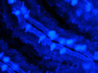
Fluorescence emission intensity from a thin section of mouse intestine stained with Alexa Fluor 350 wheat germ agglutinin, a blue fluorescent lectin that is specific to the mucus of goblet cells.
Indian Muntjac
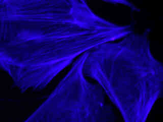
Fluorescence emission intensity from a culture of Indian Muntjac Deerskin Fibroblasts stained with Alexa Fluor 350 phalloidin, which targets actin.
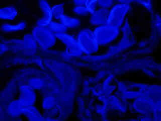
Mouse Kidney Tissue
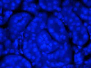
Fluorescence emission intensity from a thin section of mouse kidney stained with multiple fluorophores.
Indian Muntjac Chromosomes
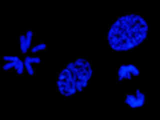
Fluorescence emission intensity from a culture of Indian Muntjac deerskin fibroblast cells adherent on a cover glass.
Contributing Authors
Related Nikon Products
Share this article:


