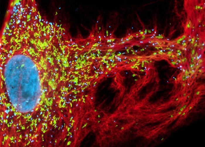
African Water Mongoose Skin Fibroblast Cells (A.P. Mongoose Line)
In a double immunofluorescence experiment, an adherent culture of APM fibroblasts was fixed, permeabilized, blocked with 10-percent normal goat serum, and then treated with a cocktail of mouse anti-vimentin and rabbit anti-PMP 70 (peroxisomal membrane protein 70) primary antibodies followed by goat anti-mouse and anti-rabbit secondary antibodies (IgG) conjugated to Alexa Fluor 647 and Alexa Fluor 750, respectively. The mitochondrial network was also targeted with MitoTracker Red CMXRos and cell nuclei were counterstained with Hoechst 33342. Images were recorded in grayscale with a 12-bit digital camera coupled to a Nikon Eclipse 80i microscope equipped with bandpass emission fluorescence filter optical blocks. During the processing stage, the image channel for Hoechst 33342 was pseudocolored with an RGB value corresponding to its fluorophore emission spectral profile, while the image channels for Alexa Fluor 750, Alexa Fluor 647, and MitoTracker Red CMXRos were pseudocolored cyan, red, and green, respectively.
Featured in:
Share this page:


