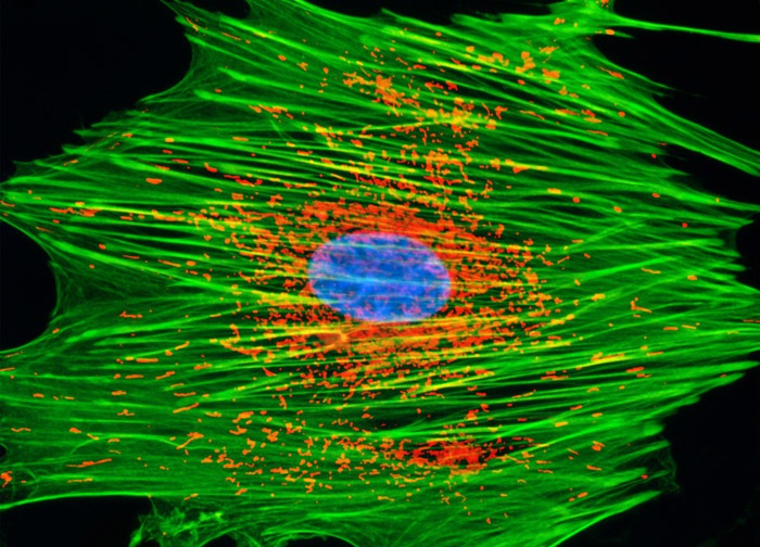
African Water Mongoose Skin Fibroblast Cells (A.P. Mongoose Line)
Using a popular triple-fluorophore staining technique for mitochondria, filamentous actin, and the nucleus, the log phase monolayer culture of A.P. Mongoose cells illustrated above was first treated with MitoTracker Red CMXRos for one hour, and then fixed with medium containing 3.7-percent paraformaldehyde. After permeabilization and blocking with bovine serum albumen, the cells were labeled with Alexa Fluor 488 conjugated to phalloidin and counterstained with Hoechst 33258. Images were recorded in grayscale with a 12-bit digital camera coupled to either a Nikon E-600 or Eclipse 80i microscope equipped with bandpass emission fluorescence filter optical blocks. During the processing stage, individual image channels were pseudocolored with RGB values corresponding to each of the fluorophore emission spectral profiles.
Featured in:
Share this page:


