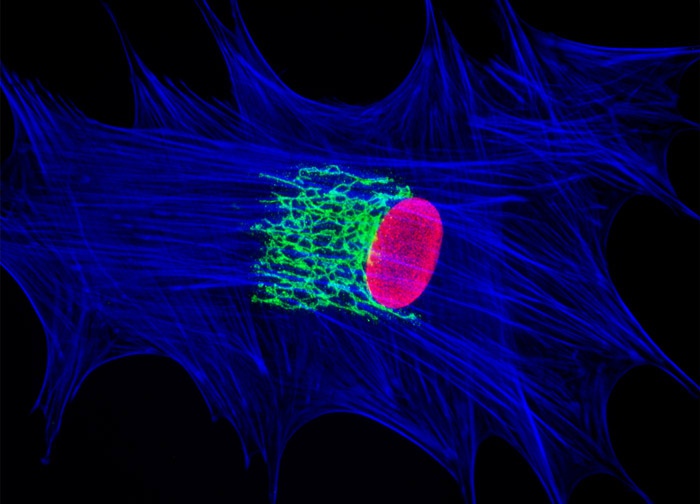
African Water Mongoose Skin Fibroblast Cells (A.P. Mongoose Line)
The close proximity between the Golgi complex and nuclei in African water mongoose cells (illustrated above) was probed in a double immunofluorescence experiment with mouse anti-NPCP (nuclear pore complex protein) and rabbit anti-giantin primary antibodies. The antibody targets were visualized with goat secondary antibodies conjugated to Alexa Fluor 568 and Alexa Fluor 488, respectively, while the actin cytoskeletal framework was labeled with Alexa Fluor 350 conjugated to phalloidin. Images were recorded in grayscale with a 12-bit digital camera coupled to either a Nikon E-600 or Eclipse 80i microscope equipped with bandpass emission fluorescence filter optical blocks. During the processing stage, individual image channels were pseudocolored with RGB values corresponding to each of the fluorophore emission spectral profiles.
Featured in:
Share this page:


