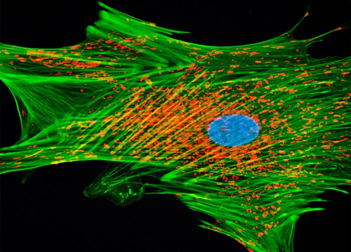
African Water Mongoose Skin Fibroblast Cells (A.P. Mongoose Line)
Applying a collection of popular mitochondrial, actin, and DNA probes, the culture of A.P. Mongoose cells presented above was grown to log phase, treated with MitoTracker Red CMXRos before fixing, and then labeled with phalloidin and Hoechst 33258 after permeabilization. The red fluorescence arises from the mitochondrial dye (MitoTracker), while the phalloidin was conjugated to Alexa Fluor 488 to generate green fluorescence. Nuclei are rendered in light blue. Images were recorded in grayscale with a 12-bit digital camera coupled to either a Nikon E-600 or Eclipse 80i microscope equipped with bandpass emission fluorescence filter optical blocks. During the processing stage, individual image channels were pseudocolored with RGB values corresponding to each of the fluorophore emission spectral profiles.
Featured in:
Share this page:


