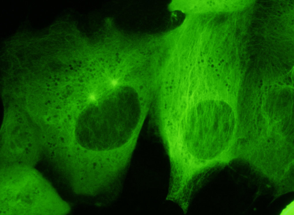
Canine Kidney Cell Tubulin
YFP Bandpass Emission (Narrow Bandwidth Excitation) Blue-Green Set
Fluorescence emission intensity from a culture of normal canine kidney epithelial (Madin-Darby; MDCK line) cells that were transfected with a pEYFP-Tubulin plasmid subcellular localization vector. Cells were transiently transfected and cultured in nutrient medium for a minimum of 24 hours before recording images. The enhanced yellow fluorescent protein gene used in these studies contains several important amino acid substitutions that shift the emission maximum of green fluorescent protein (GFP) by approximately 18 nanometers, from 509 to 527 nanometers. However, the fluorescence emission still appears green when observed in the microscope eyepieces. One of the cells (left-hand side) in the image above is entering prophase as evidenced by the separation of centrosomes above the nucleus.
Share this page:


