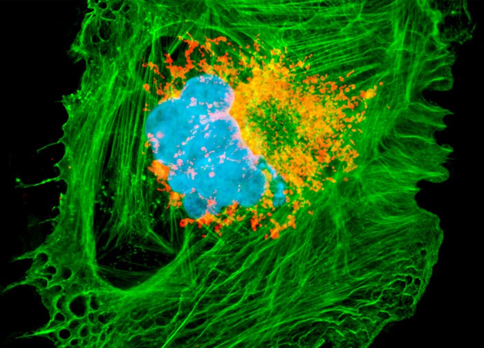
Chinese Hamster Ovary Cells (CHO-K1 Line)
MitoTracker Orange CMTM Ros was utilized to fluorescently label the active mitochondria in a culture of Chinese hamster ovary cells (illustrated above). The specimen was additionally labeled with Oregon Green 488 conjugated to phalloidin and Hoechst 33258 to visualize the intracellular F-actin network and nuclear DNA, respectively. Images were recorded in grayscale with a 12-bit digital camera coupled to either a Nikon E-600 or Eclipse 80i microscope equipped with bandpass emission fluorescence filter optical blocks. During the processing stage, individual image channels were pseudocolored with RGB values corresponding to each of the fluorophore emission spectral profiles.
Featured in:
Share this page:


