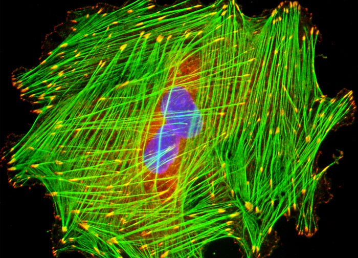
Embryonic Rat Thoracic Aorta Medial Layer Myoblast Cells (A-10 Line)
A culture of A-10 cells was immunofluorescently labeled with primary anti-vinculin mouse monoclonal antibodies followed by goat anti-mouse Fab fragments conjugated to Cy3 (red fluorescence). In addition, the specimen was simultaneously stained for DNA with the ultraviolet-absorbing probe DAPI (blue fluorescence), and for the cytoskeletal filamentous actin network with Alexa Fluor 488 (green fluorescence) conjugated to phalloidin. Images were recorded in grayscale with a 12-bit digital camera coupled to either a Nikon E-600 or Eclipse 80i microscope equipped with bandpass emission fluorescence filter optical blocks. During the processing stage, individual image channels were pseudocolored with RGB values corresponding to each of the fluorophore emission spectral profiles.
Featured in:
Share this page:


