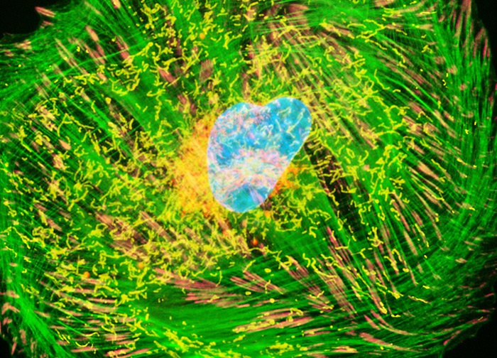
Embryonic Rat Thoracic Aorta Medial Layer Myoblast Cells (A-10 Line)
Focal adhesions and the Golgi complex were visualized in an adherent monolayer culture of A-10 cells by immunofluorescent treatment with mouse anti-vinculin primary antibodies and rabbit primary antibodies to giantin (a protein resident in the Golgi complex of mammalian cells) followed by goat anti-mouse Fab fragments conjugated to Alexa Fluor 750 and goat anti-rabbit secondary antibody fragments (heavy and light chain) conjugated to Alexa Fluor 568. The actin cytoskeletal network and mitochondria were simultaneously imaged with Alexa Fluor 488 conjugated to phalloidin and MitoTracker Deep Red 633, respectively. Nuclei were counterstained with Hoechst 33258. Images were recorded in grayscale with a 12-bit digital camera coupled to either a Nikon E-600 or Eclipse 80i microscope equipped with bandpass emission fluorescence filter optical blocks. During the processing stage, individual image channels were pseudocolored with RGB values corresponding to each of the fluorophore emission spectral profiles with the exception of MitoTracker Deep Red 633, which was pseudocolored yellow, and Alexa Fluor 750, which was pseudocolored magenta.
Featured in:
Share this page:


