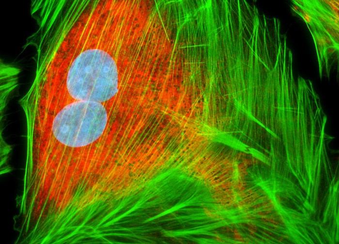
Embryonic Rat Thoracic Aorta Medial Layer Myoblast Cells (A-10 Line)
The A-10 myoblast cells presented in the digital image above were resident in an adherent culture stained for F-actin with Alexa Fluor 488 conjugated to phalloidin (green fluorescence), and for nuclear DNA with the bis-benzimidazole dye Hoechst 33258 (blue fluorescence). In addition, the culture was immunofluorescently labeled with Alexa Fluor 568 conjugated to goat secondary antibodies that target mouse anti-PDI (protein disulfide isomerase) primary antibodies (red fluorescence). Images were recorded in grayscale with a 12-bit digital camera coupled to either a Nikon E-600 or Eclipse 80i microscope equipped with bandpass emission fluorescence filter optical blocks. During the processing stage, individual image channels were pseudocolored with RGB values corresponding to each of the fluorophore emission spectral profiles.
Featured in:
Share this page:


