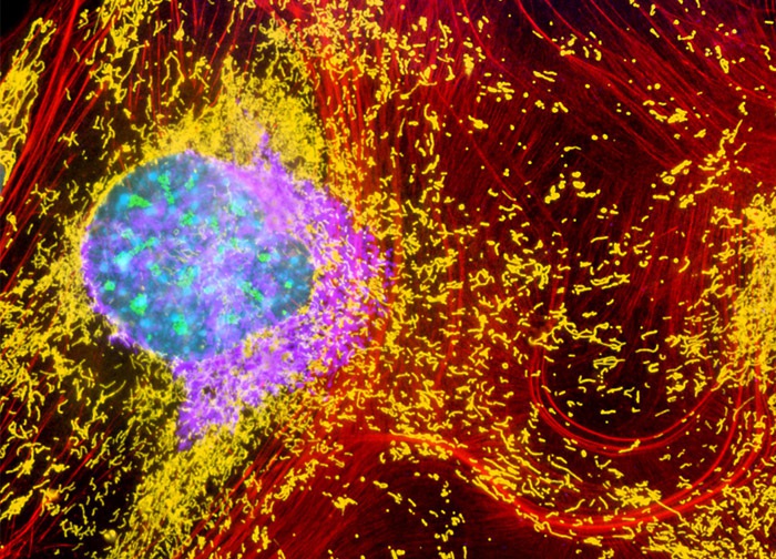
Embryonic Swiss Mouse Fibroblast Cells (3T3 Line)
The 3T3 fibroblast cell culture featured above was fixed, permeabilized, washed, and blocked with 10-percent normal goat serum in phosphate-buffered saline prior to immunofluorescent labeling with rabbit primary antibodies to giantin, a protein resident in the Golgi complex of mammalian cells, and mouse primary antibodies to fibrillarin, a component of a nucleolar small nuclear ribonucleoprotein (SnRNP). The culture was subsequently stained with a mixture of secondary antibodies conjugated to Alexa Fluor 750 to visualize the giantin and Alexa Fluor 488 (green) to visualize the fibrillarin. In addition, mitochondria were labeled with MitoTracker Deep Red 633, the filamentous actin network was counterstained with Alexa Fluor 568 (red) conjugated to phalloidin, and nuclei were targeted with Hoechst 33258 (blue). Images were recorded in grayscale with a 12-bit digital camera coupled to either a Nikon E-600 or Eclipse 80i microscope equipped with bandpass emission fluorescence filter optical blocks. During the processing stage, individual image channels were pseudocolored with RGB values corresponding to each of the fluorophore emission spectral profiles with the exception of MitoTracker Deep Red 633, which was pseudocolored yellow, and Alexa Fluor 750, which was pseudocolored purple.
Featured in:
Share this page:


