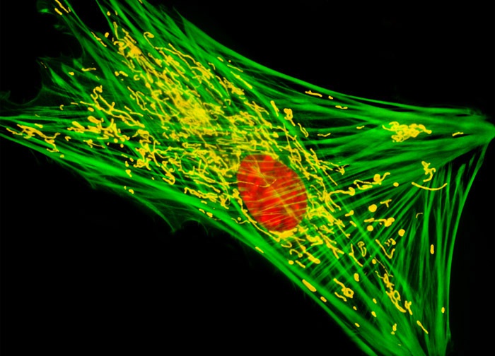
Grey Fox Lung Fibroblast Cells (FoLu Line)
A triplet of fluorophores was utilized to label the culture of FoLu cells illustrated above. Alexa Fluor 488 conjugated to phalloidin, MitoTracker Deep Red 633, and SYTOX Orange, enabled the visualization of F-actin, mitochondria, and cell nuclei, respectively. Images were recorded in grayscale with a 12-bit digital camera coupled to either a Nikon E-600 or Eclipse 80i microscope equipped with bandpass emission fluorescence filter optical blocks. During the processing stage, individual image channels were pseudocolored with RGB values corresponding to each of the fluorophore emission spectral profiles with the exception of MitoTracker Deep Red 633, which was pseudocolored yellow.
Featured in:
Share this page:


