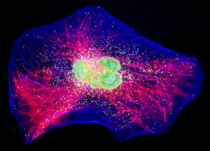
Guinea Pig Colorectal Adenocarcinoma Epithelial Cells (GPC-16 Line)
An adherent culture of GPC-16 epithelial cells (shown above) was treated with a cocktail of mouse anti-vimentin and rabbit anti-PMP 70 (peroxisomal membrane protein) primary antibodies, followed by goat anti-mouse and anti-rabbit secondary antibodies conjugated to Cy3 and Alexa Fluor 647, respectively, to target intermediate filaments and peroxisomes. The filamentous actin network was imaged with Coumarin conjugated to phalloidin and DNA in the cell nucleus was counterstained wit SYTOX Green. Images were recorded in grayscale with a 12-bit digital camera coupled to either a Nikon E-600 or Eclipse 80i microscope equipped with bandpass emission fluorescence filter optical blocks. During the processing stage, individual image channels were pseudocolored with RGB values corresponding to each of the fluorophore emission spectral profiles with the exception of Alexa Fluor 647, which was pseudocolored yellow.
Featured in:
Share this page:


