HeLa Cell Peroxisomes and Actin
FITC-Texas Red Bandpass Emission (Dual Band Excitation) Set
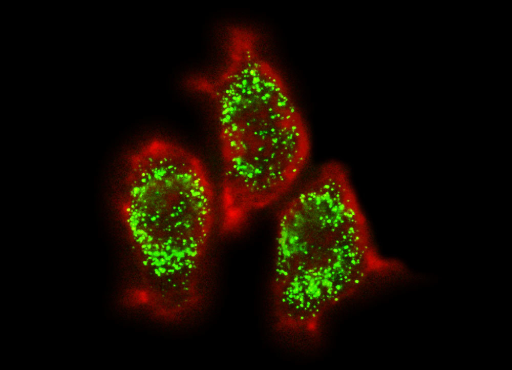
Fluorescence emission intensity from a culture of HeLa carcinoma cells transfected with an EGFP-peroxisomal targeting signal 1 (PTS1) fusion protein and stained with Alexa Fluor 546-phalloidin. These fluorescent probes target the peroxisomes and cytoskeletal actin filament network, respectively. The visible light absorption maximum of the EGFP-PTS1 chimera is 488 nanometers and the emission maximum occurs at 507 nanometers, while the corresponding values for Alexa Fluor 546 are 556 and 573 nanometers. In addition, the specimen was simultaneously stained with Hoechst 33258 (targeting the DNA in the nucleus; blue emission). Note the absence of signal from the blue fluorophore, but the bright orange-red fluorescence exhibited by the cytoskeletal actin filaments and the intense green emission from peroxisomes in the cytoplasm.
Share this page:


