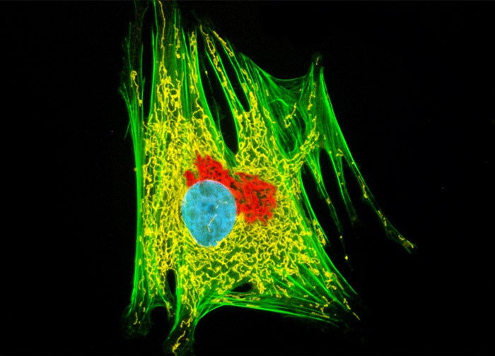
Horse Dermal Fibroblast Cells (NBL-6 Line)
Golgi bodies in a culture of horse dermal fibroblast cells (depicted above) were immunofluorescently targeted with rabbit anti-giantin primary antibodies, followed by goat anti-rabbit secondaries conjugated to Alexa Fluor 568 (yielding red emission). Mitochondria, F-actin, and nuclei present in the culture were also labeled with MitoTracker Deep Red 633, Alexa Fluor 488 conjugated to phalloidin (green emission), and Hoechst 33258, respectively. Images were recorded in grayscale with a 12-bit digital camera coupled to either a Nikon E-600 or Eclipse 80i microscope equipped with bandpass emission fluorescence filter optical blocks. During the processing stage, individual image channels were pseudocolored with RGB values corresponding to each of the fluorophore emission spectral profiles with the exception of MitoTracker Deep Red 633, which was pseudocolored yellow, and Hoechst 33258, which was pseudocolored cyan.
Featured in:
Share this page:


