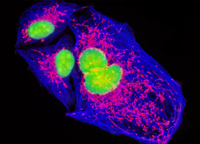
Human Bone Osteosarcoma Cells (U-2 OS Line)
Immunofluorescence, a subcellular localization vector, and a classic nucleic acid stain were utilized to triple label the culture of U-2 OS cells presented in the digital image above. The specimen was transfected with a pDsRed-Mitochondria plasmid localization vector to localize a red fluorescent protein tag to the intracellular mitochondrial network and was treated with mouse anti-alpha-tubulin followed by a secondary goat anti-mouse antibody conjugated to Marina Blue to target the microtubule network. Cell nuclei were counterstained with SYTOX Green. Images were recorded in grayscale with a 12-bit digital camera coupled to either a Nikon E-600 or Eclipse 80i microscope equipped with bandpass emission fluorescence filter optical blocks. During the processing stage, individual image channels were pseudocolored with RGB values corresponding to each of the fluorophore emission spectral profiles.
Featured in:
Share this page:


