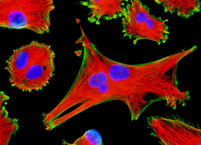
Human Brain Glioma Cells (U-118 MG Line)
The U-118 MG glioma cells presented in the digital image above were resident in a culture that was immunofluorescently labeled with anti-tubulin mouse monoclonal primary antibodies followed by goat anti-mouse Fab fragments conjugated to Texas Red. In addition, the specimen was stained with Alexa Fluor 488 conjugated to phalloidin and Hoechst 33342, targeting the cytoskeletal filamentous actin network and DNA in the cell nucleus, respectively. Images were recorded in grayscale with a 12-bit digital camera coupled to either a Nikon E-600 or Eclipse 80i microscope equipped with bandpass emission fluorescence filter optical blocks. During the processing stage, individual image channels were pseudocolored with RGB values corresponding to each of the fluorophore emission spectral profiles.
Featured in:
Share this page:


