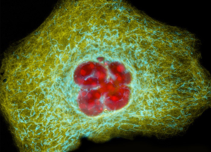
Human Cervical Adenocarcinoma Cells (HeLa Line)
A culture of HeLa cervical carcinoma cells (presented above) was simultaneously transfected with three chimeric plasmid subcellular localization vectors. DsRed2, ECFP, and EYFP plasmid vectors were utilized to localize the nucleus, mitochondria, and actin, respectively. Images were recorded in grayscale with a 12-bit digital camera coupled to either a Nikon E-600 or Eclipse 80i microscope equipped with bandpass emission fluorescence filter optical blocks. During the processing stage, individual image channels were pseudocolored with RGB values corresponding to each of the fluorophore emission spectral profiles.
Featured in:
Share this page:


