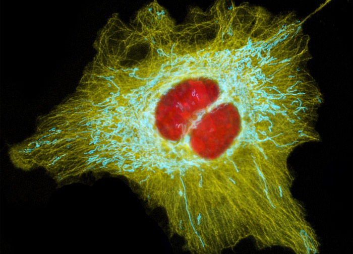
Human Cervical Adenocarcinoma Cells (HeLa Line)
Transient transfection of a log phase culture of HeLa cells (illustrated above) with multiple chimeric plasmid subcellular localization vectors (DsRed2-Nucleus, ECFP-Mitochondria, and EYFP-Tubulin) enabled the localization of a red protein tag to the cell nuclei, a cyan tag to the intracellular network of mitochondria, and a yellow tag to microtubules. Images were recorded in grayscale with a 12-bit digital camera coupled to either a Nikon E-600 or Eclipse 80i microscope equipped with bandpass emission fluorescence filter optical blocks. During the processing stage, individual image channels were pseudocolored with RGB values corresponding to each of the fluorophore emission spectral profiles.
Featured in:
Share this page:


