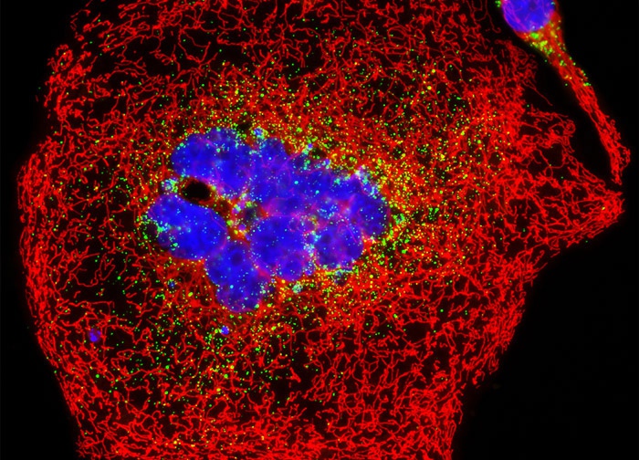
Human Cervical Adenocarcinoma Cells (HeLa Line)
The human adenocarcinoma cell culture featured in the digital image above was transfected with an EGFP-peroxisomal targeting signal 1 (PTS1) fusion protein and stained with MitoTracker Red CMXRos. These fluorescent probes target peroxisomes and mitochondria, respectively. The visible light absorption maximum of the EGFP-PTS1 chimera is 488 nanometers and the emission maximum occurs at 507 nanometers. In addition, the specimen was simultaneously stained with Hoechst 33342 (targeting the DNA in the nucleus; blue emission). Images were recorded in grayscale with a 12-bit digital camera coupled to either a Nikon E-600 or Eclipse 80i microscope equipped with bandpass emission fluorescence filter optical blocks. During the processing stage, individual image channels were pseudocolored with RGB values corresponding to each of the fluorophore emission spectral profiles.
Featured in:
Share this page:


