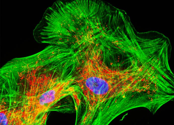
Human Cortical Neuronal Cells (HCN-1A Line)
Three fluorophores and a phallotoxin were utilized to triple label the culture of HCN-1A cells shown in the digital image above. Mitochondria were targeted with MitoTracker Red CMXRos, the cytoskeletal F-actin network was localized with Alexa Fluor 488 conjugated to phalloidin, and DNA in the cell nucleus was counterstained with Hoechst 33258. Images were recorded in grayscale with a 12-bit digital camera coupled to either a Nikon E-600 or Eclipse 80i microscope equipped with bandpass emission fluorescence filter optical blocks. During the processing stage, individual image channels were pseudocolored with RGB values corresponding to each of the fluorophore emission spectral profiles.
Featured in:
Share this page:


